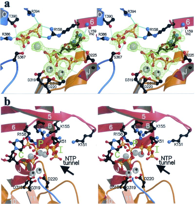
FIG. 2.
The rUTP complex. Secondary structure elements are color coded as in Fig. 1a and labeled as in Fig. 1c. (a) Close-up view of the region around the catalytic site of the enzyme (stereo view). The orientation is as in Fig. 1c, and the representation of atoms is as in Fig. 1b. Amino acids involved in binding the nucleotides are labeled. To the right is the nucleotide bound to the C site, with the uracil base hydrogen bonded to the polypeptide main chain. To the left is the tP moiety of the nucleotide bound at the P site. Hydrogen bonds are represented as in Fig. 1b. Divalent metals (Mn2+) are displayed as gray spheres; *A and *B identify the catalytic ions (see text). mc, main chain. (b) Stereo view displaying the C, P, and I sites (green) in the enzyme's active-site region in the rUTP complex. Shown is a view from the thumb. Labeling is as described in the legend to panel a. Arrow, access route of nucleotides through the NTP tunnel defined previously (8).

