
Fig. 4.
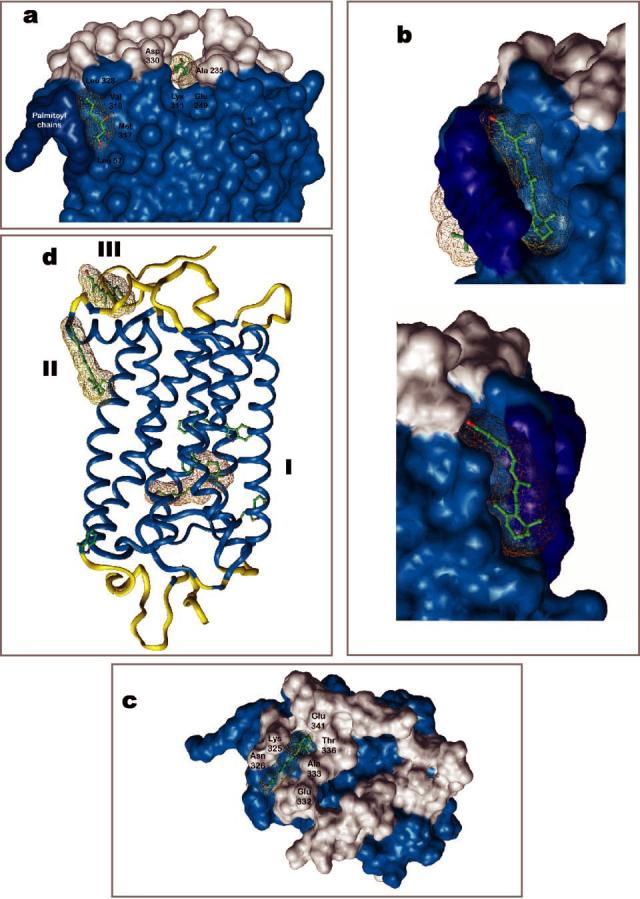
Putative binding sites of retinals on rhodopsin. Two heptane-1,2,3-triol (HTPO)-binding sites were identified in the crystal structure of rhodopsin (15, 16), one site is located close to the palmitoylation site (site II) and the second in the vicinity of C terminus and the 3rd cytoplasmic loop. Additional four HTO molecules are bound to extracellular half of rhodopsin (not shown) (a). The large site II can accommodate all-trans-retinal, 11-cis-retinal, or both together (b). Site III is located closer to the surface and could be the exit site for all-trans-retinal (c). In all figures, the transmembrane segments are colored in light blue and palmitoyl chains in dark blue. Site I is the original binding site for the chromophore, 11-cis-retinylidene, which undergoes photoisomerization. All three sites and the side chains of Trp residues are depicted in panel d.