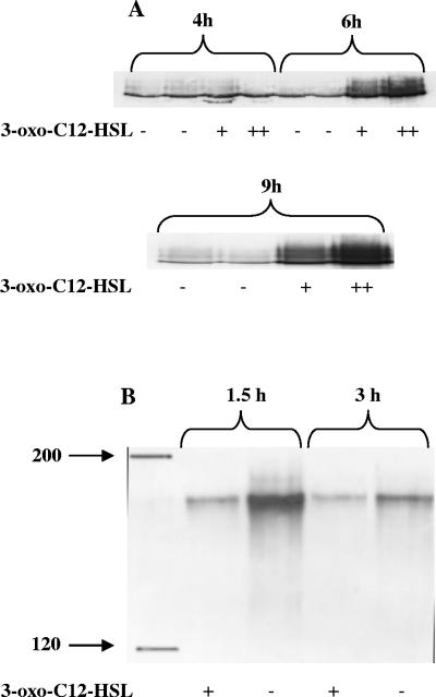
FIG. 4.
(A) Western blot of cell wall proteins of S. aureus grown for 4, 6, and 9 h in the absence of 3-oxo-C12-HSL (lanes −) or in the presence of 3-oxo-C12-HSL at concentrations of 5 μM (lanes +) and 15 μM (lanes ++) and probed with horseradish peroxidase-conjugated rabbit-anti-rat immunoglobulin G for detection of protein A. (B) Western ligand blot of cell wall proteins of S. aureus grown for 1.5 h or 3 h in the presence (lanes +) or in the absence (lanes −) of 3-oxo-C12-HSL and probed with human fibronectin. The lane on the left shows the positions of molecular mass marker proteins (in kDa).

