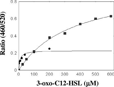
FIG. 8.
3-oxo-C12-HSL disturbs the membrane dipole potential. Changes in the dipole potential were determined spectrofluorometrically using the dipole potential fluorescent sensor di-8-ANEPPS to measure the variation in the fluorescence ratio, R(460/520), as a function of 3-oxo-C12-HSL concentration using phospholipid liposomes (▪) and S. aureus membranes •.

