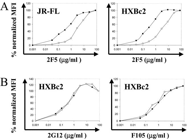
FIG. 6.
(A) Binding of the gp41 antibody 2F5 to gp160ΔCT from HXBc2 (right) and JR-FL (left) on beads without a membrane (open squares) and fully reconstituted PLs (closed squares). PLs and beads were probed with increasing concentrations of 2F5 antibody and anti-human IgG-PE antibody, respectively, and analyzed by FACS. The mean fluorescence intensity (MFI) was plotted as percent maximal MFI at the given antibody concentration. (B) Binding of the antibodies 2G12 (left) and F105 (right) to the PLs (closed squares) and beads (open squares) was performed as described for 2F5 binding above.

