FIG. 1.
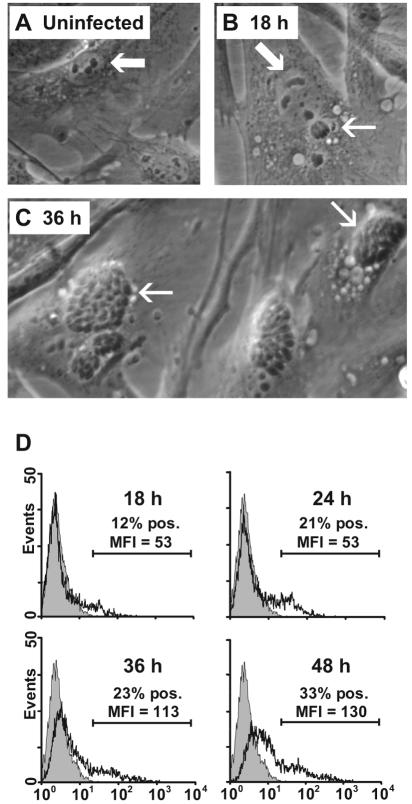
Microscopic and flow-cytometric detection of N. caninum infection of bovine primary dermal fibroblast cultures at different time points. (A) In uninfected fibroblasts, the cell morphology reflects the tissue origin with an ovoid, large, and light nucleus with evident nucleoli (arrow). (B) After 18 h of infection, a single tachyzoite within a parasitophorous vacuole (thin arrow) is seen separate from a cell nucleus (wide arrow). (C) After 36 h of infection, fibroblasts contain numerous groups of tachyzoites (arrows). (D) Flow-cytometric intracellular detection of N. caninum antigens in permeabilized fibroblasts labeled with the parasite-specific MAb 5B6 and a fluorochrome-conjugated secondary MAb (open histograms). The shaded histograms represent labeled uninfected fibroblasts; secondary MAb controls were similar (not shown). The results shown are from fibroblast culture of one animal representative of four repeated twice with similar results. MFI, mean fluorescence intensity; pos., positive.

