FIG. 3.
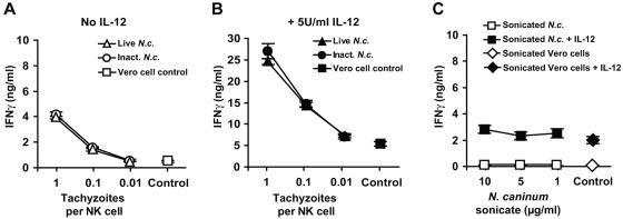
IFN-γ production from NK cells exposed to live, heat-inactivated, or sonicated N. caninum tachyzoites. (A) IFN-γ in supernatants of IL-2-activated NK cells exposed for 24 h to Percoll-purified live tachyzoites (triangles), heat-inactivated tachyzoites (56°C for 50 min; circles), or Percoll-purified uninfected Vero cell controls, corresponding to the greatest possible contamination (squares). (B) As for panel A in the presence of 5 U/ml bovine rIL-12. Note the different values on the y axis. The results shown are means ± SEM of two animals analyzed in triplicate representative of five individual experiments, in two of which inactivated tachyzoites were included. (C) IFN-γ levels in NK culture supernatants in the presence of sonicate of N. caninum at the indicated concentrations (squares) or sonicated, Percoll-purified Vero cells as controls (diamonds), in the absence (open) or presence (filled) of 5 U/ml bovine rIL-12. The results shown are means ± SEM of four animals analyzed in triplicate representative of two individual experiments.

