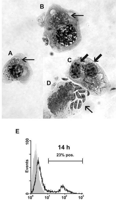
FIG. 6.
Giemsa-stained cytospin preparation of N. caninum-infected bovine NK cells. (A) The arrow points at two tachyzoites in a parasitic vacuole. (B) NK cell harboring several tachyzoites (arrow). (C) Binucleated N. caninum-infected NK cell. The arrows point at the two nuclei. (D) Free N. caninum tachyzoites (arrow) emerging from lysed NK cell. (E) Flow-cytometric histogram of infected NK cells permeabilized and labeled with the parasite-specific MAb 5B6 and a fluorochrome-conjugated secondary MAb. The shaded histograms represent labeled uninfected NK cells; secondary controls were similar (not shown). The results shown are from NK cell cultures of one animal representative of two; the experiment was repeated twice with similar results. pos., positive.

