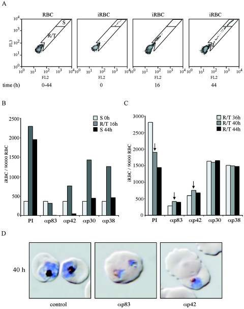
FIG. 5.
Time course of inhibition of parasite replication in the presence of anti-MSP-1 antibodies. Synchronized schizont cultures were assayed with rabbit sera specific for the four MSP-1D processing fragments p83, p30, p38, and p42. The numbers of iRBCs and their relative DNA content were determined at 4-h intervals by flow cytometry. (A) Overview of the experimental approach. Left to right: flow cytometric pattern of uninfected RBCs; infected, synchronized RBC cultures (iRBC) showing the position of schizonts (gate S) at time zero; iRBCs at 16 h, demonstrating the position of ring stage parasites and early trophozoites (gate R/T); iRBCs at 44 h, where a substantial fraction is again in schizont stage while, due to the diminishing synchronization of the culture, late trophozoites are still present as new ring stage parasites reappear. The latter ring stage parasites are not seen when full inhibition by antibodies prevents the transition from intact trophozoites to schizonts (see below). (B) Time course of parasite multiplication in the presence of preimmune serum (PI) or of serum raised against (α) p83, p42, p30, or p38, as indicated; iRBCs per 90,000 RBCs were quantified at different time points by using the gates as shown in panel A. Light-grey columns, starting cultures containing exclusively schizonts (S); medium-grey columns, ring stage parasites and early trophozoites (R/T) at 16 h; dark-grey columns, schizonts (S) at 44 h. (C) Fates of ring stage parasites and trophozoites at different time points in the presence of preimmune serum (PI) or of sera raised against the four MSP-1 subunits as indicated. Light-grey, medium-grey, and dark-grey columns show the numbers of iRBCs per 90,000 RBCs at 36, 40, and 44 h. Arrows indicate the cultures from which the microscopic analyses shown in panel D are taken. (D) Microscopy of parasites cultured for 40 h in the absence and presence of inhibiting antibodies. Samples were taken from the experiment represented by panels B and C, as indicated by arrows in panel C. Parasites were stained with Giemsa. Early schizonts are detected in the presence of preimmune serum (control), whereas typical crisis forms which have stopped development are seen in the presence of anti-p83 (αp83) and anti-p42 (αp42) serum. The morphologies shown are representative of the parasites detected under the conditions described.

