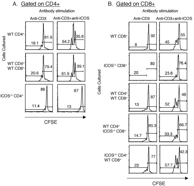
FIG. 6.
Anti-ICOS mAb can costimulate CD8 T cells in the absence of CD4 T-cell help. CD4 or CD8 T cells were isolated from wild type and ICOS−/− naïve mice, labeled with CFSE, and plated onto antibody-coated wells as indicated at the top. The purity of the CD4 and CD8 T cells was routinely 99% or more. (A) Division of CFSE-labeled CD4 T cells. Cells were stimulated with either anti-CD3 plus hamster IgG or with anti-CD3 plus anti-ICOS, as indicated at the top. Unstimulated cells showed no cell division. All events shown were gated on live CD4+ cells. The T cells used in the culture are indicated on the left. The graphs show CFSE loss in WT or ICOS−/− CD4 T cells cultured under the conditions indicated at the top. The numbers on the histograms indicate the percentages of cells in the undivided or divided populations. (B) Division of CFSE-labeled CD8 T cells alone (top panels) or in the presence of CD4 T cells from wild-type or ICOS−/− mice, as indicated on the left. T cells were stimulated with anti-CD3 plus hamster IgG or anti-CD3 plus anti-ICOS, as indicated at the top. All events were gated on live CD8+ cells. The numbers are the percentages of cells which have or have not divided. The results of this experiment are representative of two similar experiments analyzed at 48 h. The data were also analyzed at 72 h, with similar results.

