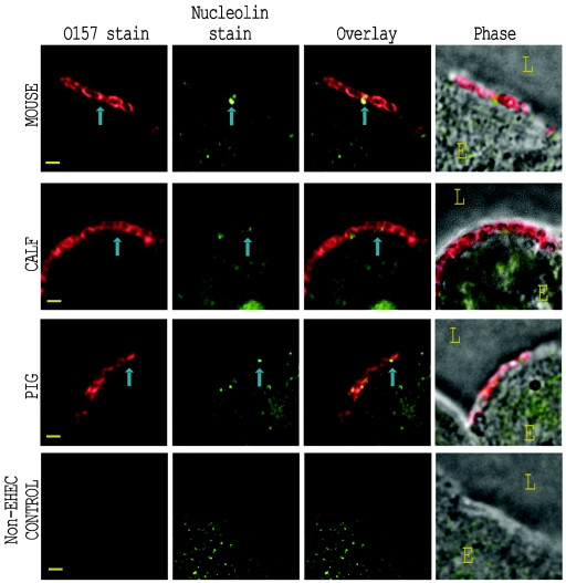
FIG. 4.
Nucleolin localization with EHEC O157:H7, detected by immunofluorescence on infected tissue sections. Sections of EHEC O157:H7 Strr-infected intestine corresponding to those depicted in Fig. 3 were immunostained with anti-nucleolin and anti-O157 sera. Representative images from EHEC O157:H7-infected mouse (top row), calf (second row), and pig (third row) are shown for stained O157 (first column) and stained nucleolin (second column). Images in the bottom row were taken from a piglet infected with the normal enteric E. coli strain 123. These fluorescent images were overlaid (third column) to demonstrate immunostained nucleolin around the site of bacterial adherence. Phase-contrast micrographs overlaid with the combined fluorescence (fourth column) depict the orientation of the epithelial cell (E) and lumen (L). Representative adherent bacteria and associated immunostained nucleolin are indicated by blue arrows. Bar, 1 μm.

