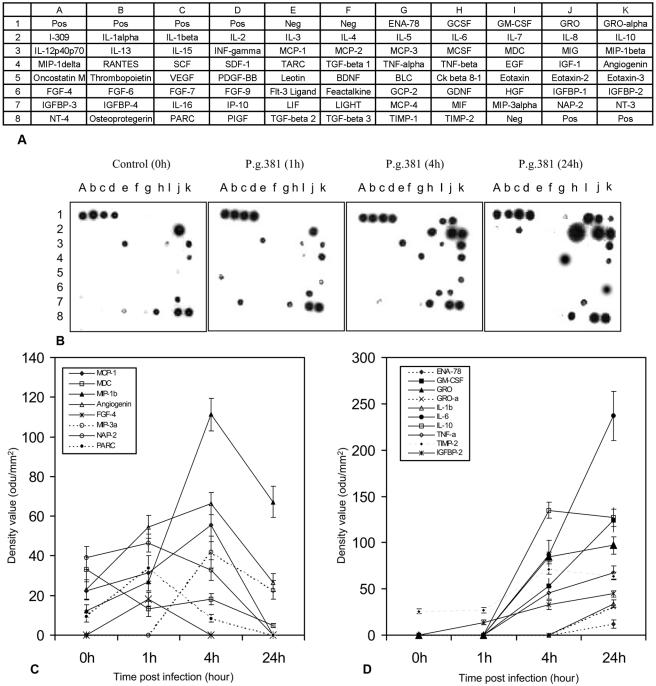
FIG. 1.
Cytokines induced by P. gingivalis in freshly elutriated human peripheral blood monocytes. Monocytes were cultured to enable differentiation into macrophages and then were exposed to live P. gingivalis bacteria at an MOI of 25:1 for 1 h, 4 h, or 24 h. Untreated cell cultures served as controls (0 h). Cell culture supernatants were subjected to a cytokine antibody array. (A) The layout shows the locations of each antibody in the array membrane. (B) The cytokine array image shows the results of one of two independent experiments obtained by exposure of membranes to X-ray film. (C and D) Average net optical intensities (mean ± standard deviation; n = 3) in optical density units (odu) for each cytokine spot induced by P. gingivalis at the indicated time points. P.g., P. gingivalis.

