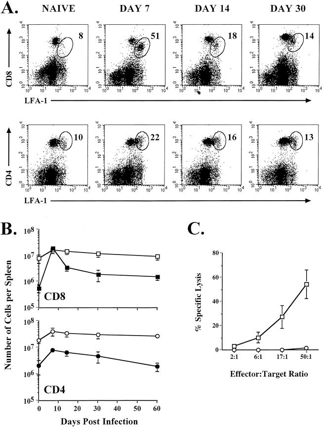
FIG. 1.
Activation and expansion of T cells following VV infection. (A) BALB/c mice were infected with VV and analyzed on the indicated days after infection by staining splenocytes with CD8α, CD4, and CD11a MAbs. The percentages of the CD8 or CD4 T-cell populations that were LFA-1hi are indicated in the upper right corners of the corresponding panels. (B) Total numbers of CD8 and CD4 T cells in the spleen that were LFA-1hi (closed symbols) or LFA-1lo (open symbols) were determined. (C) Direct ex vivo CTL activity was measured on day 7 postinfection. Killing was assayed on 51Cr-labeled uninfected (○) or VV-infected (□) target cells. Error bars indicate standard deviations.

