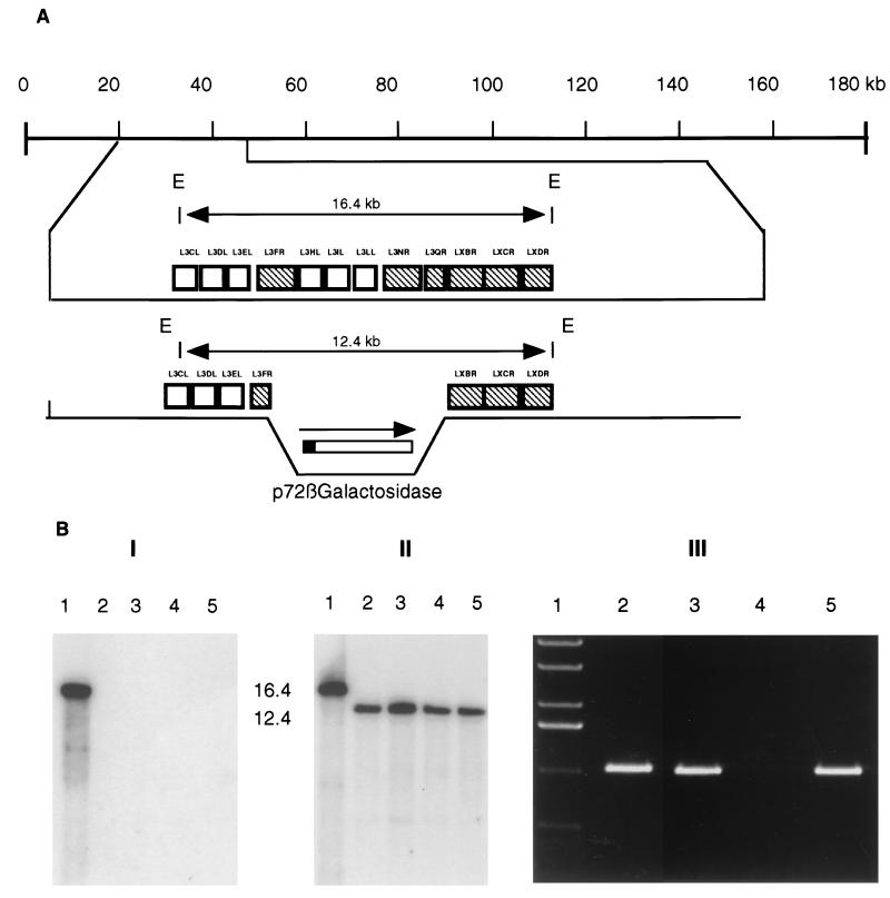
FIG. 4.
Characterization of MalΔSVD recombinants. (A) Diagram of the swine virulence determinant gene region in the parental MalΔNL isolate and the swine virulence determinant deletion mutant virus MalΔSVD. E denotes EcoRI restriction endonuclease site. (B) Southern blot analysis of parental virus (lane 1) and four independently isolated MalΔSVD viruses (lanes 2, 3, 4, and 5). Purified viral DNA was digested with EcoRI, electrophoresed, blotted, and hybridized with 32P-labeled L3IL gene probe (panel I) or 32P-labeled probe generated from the recombination transfer vector (panel II). Molecular size markers are in kilobase pairs at the left of panel ll. Panel III, PCR analysis of MalΔSVD (lanes 2 and 4) and parental MalΔNL (lanes 3 and 5) for L3IL gene sequences (lanes 4 and 5). Positive-control PCR for the MalΔSVD viral DNAs using primers for a region of the genome immediately flanking the swine virulence determinant region are included (lanes 2 and 3). Molecular size markers are shown in lane 1.

