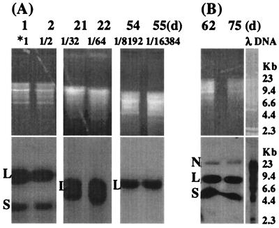
FIG. 3.
(A) BPV-1 DNA detected by Southern blot in a BPV-infected S. cerevisiae culture over 55 days. S. cerevisiae cells (10 ml; 5 × 10⁷cells/ml) infected with 0.6 μg of BPV-1 was cultured at 28°C in the dark. At 1 day, 5 ml of S. cerevisiae cells was collected for Hirt DNA preparation, with 5 ml of fresh medium added for continuous culture. This was repeated every day until 5 days. At 5 days, 5 ml of S. cerevisiae cells was held at 4°C for 15 days. Then 5 ml of fresh medium was added, and cells were cultured at 28°C. From day 21 to day 25, 5 ml of S. cerevisiae culture was collected for Hirt DNA and 5 ml of fresh medium was added every day. A total of 5 ml of the 25-day S. cerevisiae culture was held at 4°C for 25 days. Fresh medium (5 ml) was added, and cells were cultured at 28°C, diluted, and sampled as previously from 51 days to 55 days. Hirt DNA (20 μl) was resuspended in 200 μl of TE buffer, partially (days 1, 2, 21, and 22) or completely (days 54 and 55) digested with HindIII, electrophoresed on a 1% agarose gel, and blotted onto a nylon membrane. *1 indicates that the Hirt DNA contained the viral DNA from 0.03 μg of BPV-1 virus. Viral DNA dilution at the different time points is shown above each lane. The time point and effective culture dilution are shown above each lane. (B) BPV-1 DNA detected by Southern blot in a BPV-infected S. cerevisiae culture over 75 days. A total of 2 ml of S. cerevisiae cells (5 × 10⁷cells/ml) infected with 0.06 μg of BPV-1 was cultured at 28°C in the dark. Then 2 ml of fresh medium was added once a week for continuous culture. Hirt DNA prepared from S. cerevisiae infected with BPV-1 virus 62 and 75 days postinfection was electrophoresed on a 1% agarose gel without enzyme digestion. All blots were hybridized with BPV-1 DNA using [α-32P]dCTP at 3,000 Ci/mmol and exposed using Kodak BioMax film at −70°C for 24 h.

