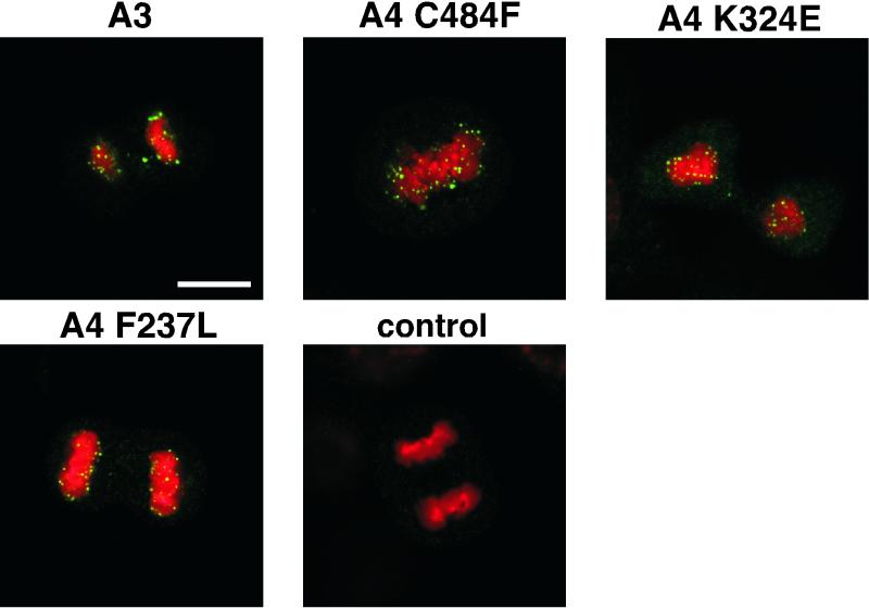
FIG. 1.
E2 protein is localized to mitotic chromosomes in suppressor mutant-transformed cells. Cells stably transformed with BPV-1 genomes of different genotypes were obtained via coselection with a Neor marker, and E2 proteins were detected by confocal microscopy using a Leica TCSNT confocal laser scanning imaging system. Green, E2 immunofluorescence with monoclonal antibody B201; red, DAPI staining of chromosomal DNA. For the confocal processing, we chose red for the DAPI signal. The upper left panel shows an anaphase mitotic cell from C127 cells transformed with the A3 mutant of BPV. The other panels show E1 suppressor mutants of the A4 genome of BPV at different stages of mitosis (E2-A4/E1 C484F, metaphase plate; E2-A4/E1 F237L, anaphase; and E2-A4/E1 324E, telophase). Mitotic figures from the nontransformed C127 cells did not stain for E2 protein (control). Bar, 10 μm. The E2-A4 mutant genome does not stably transform cells, and the localization of E2 for this allele from intact plasmids can only be assessed through transient assays and has been described previously (11); the protein in the genome context is not associated with mitotic chromosomes.

