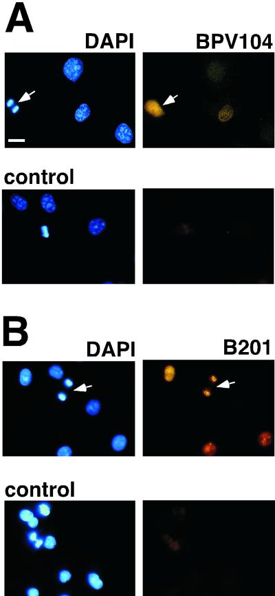
FIG. 3.
PAVA-E2 and PAVA-E1 expressed protein is detected in COS-7 cells by immunofluorescence. COS-7 cells were grown on cover slips and incubated with 5 MOI of virus for 24 h. After fixation, E2 and E1 were detected by immunofluorescence with a Zeiss axioplan fluorescence microscope using monoclonal antibodies B201 and BPV104, respectively, and the DNA was stained with DAPI. As a negative control, mock-infected cells were incubated with B201 or BPV104, followed by the Cy3-conjugated secondary antibody. Bar, 20 μm.

