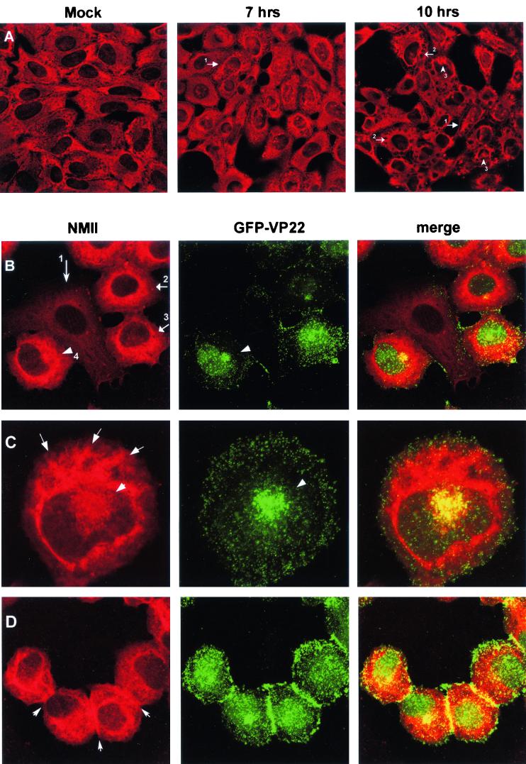
FIG.3.
Alterations in nonmuscle myosin II localization patterns in infected cells. (A) Nonmuscle myosin II retracts from the cell edge during virus infection. Mock-infected (Mock) or HSV-1-infected Vero cells were fixed and stained with antibody against nonmuscle myosin II at 7 and 10 h p.i. as indicated. Nonmuscle myosin II staining was reduced at the cell periphery (no. 1 arrows) and was accompanied by condensation into a spoke-like pattern (no. 2 arrows) and frequent perinuclear clustering (no. 3 arrows). (B to D) Alterations in nonmuscle myosin II localization patterns in infected cells. Vero cells were infected with HSV-1 166v which expressed GFP-VP22, fixed, and stained, and the distribution of anti-myosin II (red) or GFP-VP22 (green) was examined. Merged images are shown in the right panel. Arrowheads in panel B indicate perinuclear clusters. Arrow no. 1 indicates a cell not yet expressing GFP-VP22, in which NMIIA exhibits the more typical cytoplasmic staining; arrows 2 to 4 indicate cells expressing GFP-VP22, in which NMIIA staining has become more intense. Arrows in panel C indicate the condensed spoke-like pattern of nonmuscle myosin II in infected cells. Arrows in panel D indicate accumulation of NMIIA at cell-cell junctions. Several features of the alteration in nonmuscle myosin II and GFP-VP22 are shown in each of the panels as discussed in the text.

