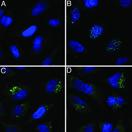
Fig. 3.
Microscopic analysis of HNP-1 and HD-5 inhibition. HeLa cells were mock treated (A) or treated with BrdUrd-labeled HPV16 PsV (B-D) in the presence of HNP-1 (C) or HD-5 (D). At 24 h after virus inoculation, the cells were stained for BrdUrd to reveal uncoated viral DNA (green). Cells were counter-stained with the DNA stain DAPI (blue).

