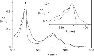
Fig. 1.
UV-visible difference absorption spectra obtained upon electrochemical reduction of 16+ (solid line) and 26+ (dashed line) at –0.50 V vs. SCE in a spectroelectrochemical thin-layer cell (5.0 × 10⁻⁴ mol·liter–1 acetonitrile solution at room temperature). The spectra were normalized to the intense band peaking at 392 nm for 16+.(Inset) Detail of the spectra in the 370–405 nm region.

