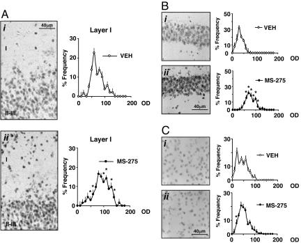
Fig. 5.
MS-275 elicits an increase of neuronal Ac-H3 immunoreactivity in the piriform cortex and hippocampus but not in the striatum. (A Left) Typical Ac-H3 immunostaining in piriform cortex neurons (layers I and II-III) of mice treated with VEH (i) or MS-275 (15 μmol/kg) (ii). (Right) Densitometric distribution profiles of Ac-H3-immunopositive neurons in layer I: VEH-(open circles) and MS-275-treated mice (solid circles). (B Left) Typical immunostaining in CA1 hippocampal neurons of mice injected with VEH (i) or MS-275 (120 μmol/kg) (ii). (Right) Densitometric distribution profiles of Ac-H3-immunopositive neurons: VEH-(open circles) and MS-275-(solid circles) treated mice. (C Left) Typical Ac-H3 immunostaining in the striatal neurons of mice injected with VEH (i) or MS-275 (120 μmol/kg) (ii). (Right) Densitometric distribution profiles of Ac-H3-immunopositive neurons: VEH-(open circles) and MS-275-(solid circles) treated mice. OD, optical density of Ac-H3 immunostaining in each cell with a 256 gray scale used as a reference. For the densitometric distribution profiles, each point is the mean ± SE of three mice. Mice were injected s.c. 2 h before perfusion. * denotes statistically significant differences between VEH- and MS-275-treated mice (P < 0.05) when data were analyzed by two-way repeated measures ANOVA followed by Student-Newman-Keuls multiple comparison.

