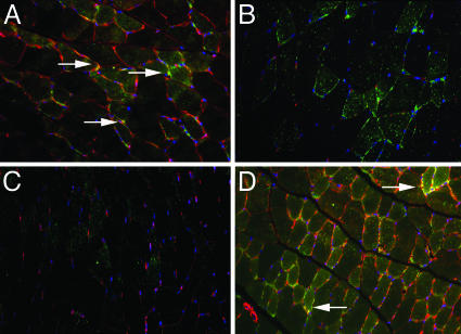
Fig. 5.
Colocalization of GLUT4 and caveolin-1 on skeletal muscle membranes of WT, ERβ-/-,ERα-/-, and ArKO mice by immunofluorescence. Muscle was stained for GLUT4 with FITC (green) and caveolin-1 with Cy3 (red). Additionally, nuclei were stained with DAPI (blue). Double staining revealed the colocalization of these proteins in WT (A) and ArKO (D) mice on the plasma membrane. However, the colocalization was never observed in ERβ-/- (B) or ERα-/- (C) mice. Arrows show the colocalization of the two receptors (yellow).

