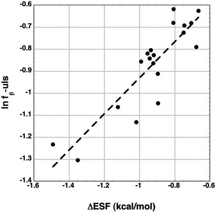
Fig. 4.
The y axis refers to the fraction of residues in the coil library β basin, which is divided by the number of residues in the upper left strip of the φ,ψ map. The logarithm of this fraction is plotted against the difference in ESF between the PII and β backbone conformations, ESF(PII) – ESF(β), for dipeptides (R = 0.88).

