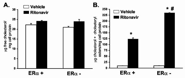
Figure 6.
Peritoneal macrophages isolated from ERα- mice accumulate more cholesteryl esters when exposed to ritonavir in vitro. Peritoneal macrophages were isolated by saline lavage and grown in culture for twenty-four hours in the presence of ritonavir (30 ng/ml) or vehicle (0.01% ethanol) plus AgLDL (50μg/ml). After twenty four hours, cell lysates were prepared and assayed for free and total cholesterol by gas chromatography. There were no significant differences in free cholesterol (Figure 6A). Total cholesterol (free cholesterol plus cholesteryl esters) dramatically increased following ritonavir treatment (Figure 6B). This increase was greater in peritoneal macrophages lacking the ERα. Bars represent the mean +/- SEM, n=4. * = significantly different from vehicle, p<0.001. # = significantly different from ERα + macrophages treated with ritonavir, p<0.001.

