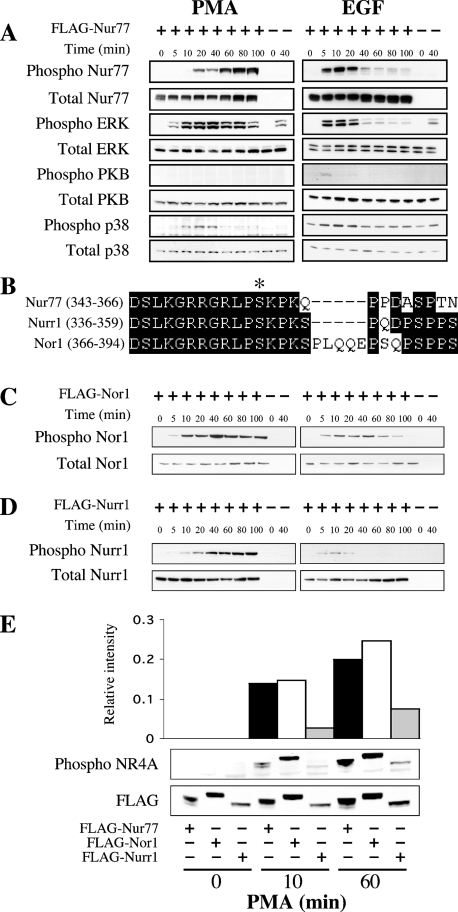
Figure 3. Nur77 Ser354 phosphorylation is stimulated in vivo by PMA and EGF.
(A) HEK-293 cells were transfected with an expression plasmid for FLAG–Nur77. Cells were then starved for 16 h and stimulated with either 400 ng/ml PMA or 100 ng/ml EGF for the times indicated. Cells were then lysed and 30 μg of soluble protein lysate was run on 4–12% gradient polyacrylamide gels. The levels of phospho-Ser354 Nur77, total Nur77, phospho-ERK1/2, total ERK1/2, phospho-Thr308 PKB, total PKB, phospho p38 and total p38 were then examined by immunoblotting. (B) Sequence alignment of murine Nur77 (GenBank® accession no. P12813), Nurr1 (GenBank® accession no. A46225) and Nor1 (Genbank® accession no. Q9QZB6) around the RSK phosphorylation site. (C) HEK-293 cells were transfected with an expression plasmid for FLAG–Nor1. Cells were then starved for 16 h and stimulated with either 400 ng/ml PMA or 100 ng/ml EGF for the times indicated. Cells were then lysed and 30 μg of soluble protein lysate was run on 4–12% gradient polyacrylamide gels. The levels of phospho-Ser377 Nor1 and total Nor1 were determined by immunoblotting. (D) HEK-293 cells were transfected with an expression plasmid for FLAG–Nurr1. Cells were then starved for 16 h and stimulated with either 400 ng/ml PMA or 100 ng/ml EGF for the times indicated. Cells were then lysed and 30 μg of soluble protein lysate was run on 4–12% gradient polyacrylamide gels. The levels of phospho-Ser347 Nurr1 and total Nurr1 were determined by immunoblotting. (E) Samples from unstimulated or PMA-stimulated cells transfected with FLAG–Nur77, FLAG–Nor1 or FLAG–Nurr1 were run on 4–12% gradient polyacrylamide gels and immunoblotted using either an anti-FLAG antibody or the anti phospho-Ser354 Nur77 antibody. Blots were visualized using fluorescent labelled secondary antibodies and the signal was quantified using a Licor Odyssey scanner. The level of the phospho signal was then calculated relative to the signal for FLAG.

