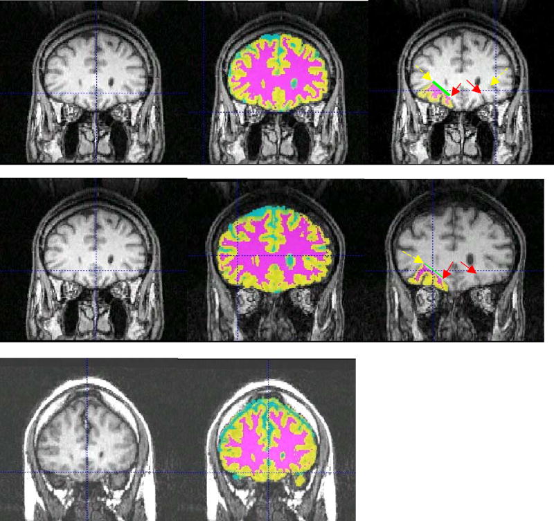
Figure 1.
For each panel, let is raw image, middle is segmented image overlaid on raw image, and right is segmented OFC overlaid on raw image.. Left side of image is right side of brain, superior is up. For segmented images: Cyan = CSF, Yellow = Gray Matter, Magenta = White Matter. The medial borders of the OFC (deepest aspect of orbital sulcus) are indicated with red (solid) arrow, whereas the lateral borders (deepest aspect of anterior horizontal ramus [AHR] of the central suclus) are indicated with yellow (dashed) arrow. The medial boundary of the white matter is indicated by the green (thick) line. Top row: image on which both AHRs are visible. Middle row: case in which one AHR is visible. Bottom row: case in which neither AHR is visible (no OFCs were drawn).

