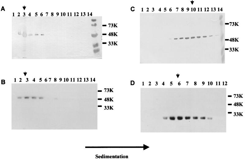
FIG. 3.
Analysis of the presence of capsid protein in fractions of iodixanol and sucrose gradients. The supernatant of infected cells (3 × 10⁶ cells/ml in 200 ml) was sedimented and separated through sucrose or iodixanol gradients. The figure shows Western blotting analysis of fractions from iodixanol (A and B) and sucrose (C and D) gradients. The fractions shown to contain capsid protein were pooled, and the sucrose or iodixanol was removed by dilution in phosphate buffer and sedimentation. (A) Full-length NV capsid, (B) NT20, (C) ID375, and (D) CT230. Sizes are shown in kilodaltons. The arrowhead shows the peak fraction in each gradient.

