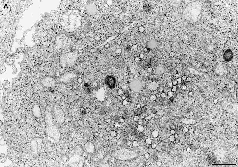
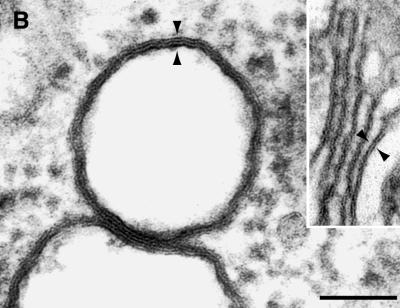
FIG. 5.
EM analysis of membrane alterations in MHV A59-infected cells. (A) Electron micrograph showing multiple DMVs in the cytoplasm of MHV-infected HeLa-MHVR cells at 5 hpi. Bar, 1 μm. (B) DMVs seen in MHV-infected 17Cl-1 cells at 7 hpi. The double membrane is fused into a trilayer. Inset: For comparison, Golgi apparatus composed of a bilayer membrane. Arrowheads indicate the thickness of the membranes. Bar, 100 nm.


