
Figure 1.
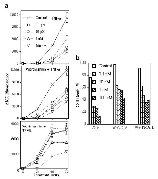
Effects of DHT on caspase activity (a) and cell death (b) in LNCaP after treatment with TNF-α and TRAIL. Caspase activity was measured in living cells with the fluorogenic substrate Ac-DEVD-AMC. The substrate (20 μm final concentration) was mixed with cells in growth medium, cells (7000/well/100 μl) were plated on 96-well plates and incubated with the substrate for 24 h to make cells adherent. Cells were then treated with DHT at indicated concentrations, and 1 h later, cells were treated with TNF-α (20 ng/ml), wortmannin (1 μm) plus TNF-α or wortmannin plus TRAIL (100 ng/ml). Substrate hydrolysis was monitored using a fluorescence reading system set to 360 nm for excitation and 460 nm for emission. Cell death was estimated by the calcein AM assay after 72 h of treatment. Each point or column represents mean values of four replicates in one of two experiments, which all gave similar results