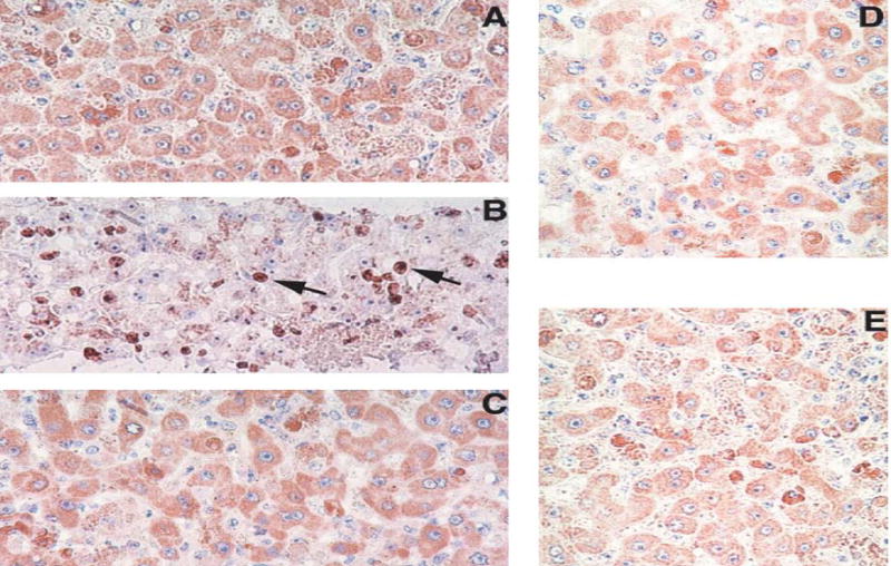
Fig. 2.

Ki-67 staining of liver sections from rhesus macaques infected with LCMV. A, B, C: Liver biopsies taken from monkey WE-ig7b before infection (A), four weeks after infection (B), and four weeks after challenge (C). D and E: Ki-67 staining of biopsy liver samples taken from ARM-iv3 (D) and ARM-ig8 (E) monkeys at four weeks after infection.
Magnifications: ×300. Note brown staining of Ki-67-positive nuclei (arrowed) in B
