
Fig 1.
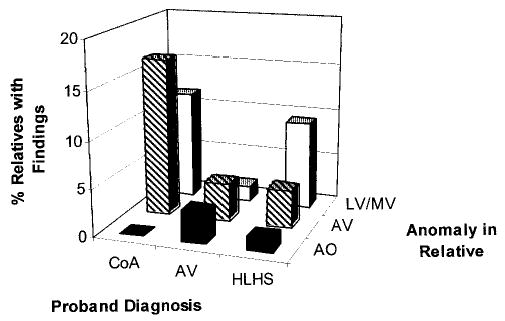
Percentage of first-degree relatives with anomaly on echocardiography according to anatomic location of defect and proband diagnosis. Anomalies classified into each anatomic group are AO (aorta: aortic dilation; n = 5), AV (aortic valve: aortic valve thickening, aortic regurgitation, bicuspid aortic valve; n = 21), LV (left ventricle: segmental thickening of the interventricular septum with protrusion into the left ventricular outflow tract, septal hypertrophy with septal to free wall ratio > 1.4; n = 10), and MV (mitral valve: mitral valve thickening, mitral regurgitation, redundant chordae tendinae; n = 9). Proband diagnoses are AVS, CoA, and HLHS.