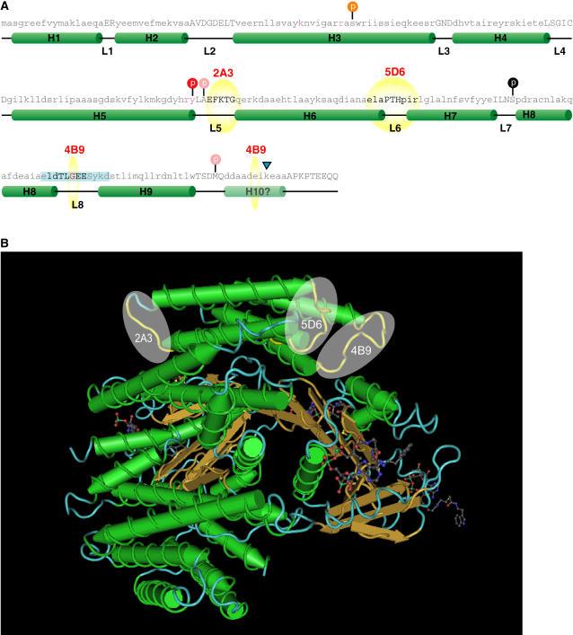
Figure 4.
GBC mAb epitopes map to loops residing on a common surface on 14-3-3s. A, Primary and secondary structural elements of GBC mAb epitopes on Arabidopsis 14-3-3 ω. The sequence of GF14ω was superimposed on the secondary structural elements from the crystal structures of tobacco and human 14-3-3s based on sequence alignment. Helices are represented by green cylinders with the helix number (Hn where n = 1–10) and loops as black lines with the designation L (loop number). The mapped GBC mAb epitopes are indicated by yellow spheres and bold text. The location of potential posttranslational events based upon the 14-3-3 literature and using sequence alignment is represented by a colored circle on top of a vertical black line. Red circles indicate a decrease in binding with the modification, while orange implies decrease in dimerization. Pink circles indicate that the specific residue is required for the modification and is not conserved in plant 14-3-3s. The residues important for phospho-Ser target ligand coordination are in red. Tryptic cleavage of GF14ω is indicated by the blue triangle. B, The location of the mAb epitopes cluster on the 14-3-3 three-dimensional structure. Using the x-ray diffraction derived three-dimensional model of the human 14-3-3 ζ complexed with AANAT PDB accession number 1IB1, the regions corresponding to the determined GBC mAb epitopes were mapped and are highlighted in yellow. The regions corresponding to mAb epitopes are represented as yellow spheres.

