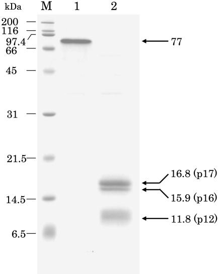
Figure 4.
SDS-PAGE on 15% polyacrylamide gel of purified PPD (type 2) from radish cotyledons. Peptides were stained by Coomassie Brilliant Blue R-250. In the nonheated sample (lane 1), only one band was stained. After heating at 95°C for 3 min (lane 2), PPD was separated into three bands. Numerals under kD denote the molecular mass of marker proteins. Lane M, Molecular markers.

