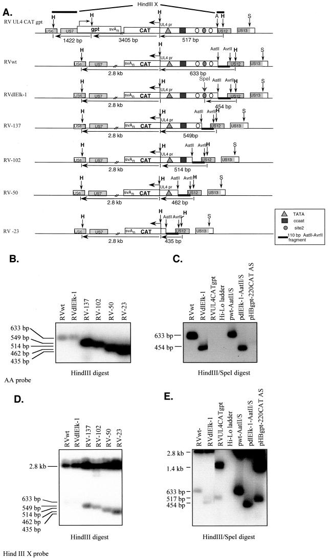
FIG.2.
Southern blot analysis of recombinant viruses. (A) Schematic genome maps of RVUL4CATgpt, RVwt, RVdlElk-1, RV-137, RV-102, RV-50, and RV-23. The UL4-CAT in its ectopic location is oriented according to the prototype position of the US genes, where transcription is from right to left. The sizes of the DNA fragments resulting from HindIII and SpeI restriction endonuclease digestion are indicated in base pairs. The genes involved in homologous recombination in shuttle vectors are shown in shaded boxes. A, S, and H represent restriction endonuclease sites AvrII, SacI, and HindIII, respectively. (B to E) Autoradiograms of Southern blots to identify the recombinant viruses by using either the 32P-labeled 110-bp AatII-Avr II DNA (AA probe [B and C]) or HindIII X probe (D and E). Lanes containing viral DNA fragments from different recombinant viruses were spliced together from the same gel. Shuttle vectors pwt-AatII/S, pdlElk-1-AatII/S, and pHBgpt-220CAT AS were used as controls. Panels D and E result from stripping the AA probe from panels B and C, respectively, and then hybridization with the HindIII X probe.

