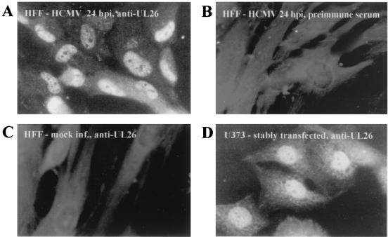
FIG. 7.
Immunofluorescence analysis of HCMV-infected HFFs and stably transfected U373 cells. Specific antiserum was used for the staining of HCMV-infected (24 hpi) or mock-infected HFFs (A and C, respectively) or stably transfected U373 cells (D). No specific signal could be observed when HCMV-infected HFFs (24 hpi) were probed with the preimmune serum (B).

