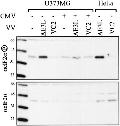
FIG. 5.
eIF2α phosphorylation after CMV and VV infection. U373MG cells were mock infected (−) or infected with CMV (+). After 24 h, the cells were mock infected (−) or infected with the indicate VV, and then lysates, prepared 24 h later, along with control extracts from VV-infected HeLa cells, were separated by SDS-PAGE and analyzed by immunoblot assay by using antibody specific for phosphorylated eIF2α (top) or total eIF2α (bottom). Molecular size markers are shown in kilodaltons to the left of each panel.

