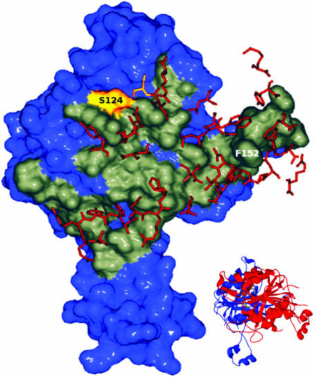
Figure 5.
Dimerization interface of RsrI MTase. One monomer of M.RsrI is shown in surface representation with the dimer interface highlighted in gray. Residues within 4 Å of this interface coming from the opposite monomer are shown as orange sticks. The S124 residue is colored yellow. The figure was created using Swiss PDB Viewer (34) and POV-RAY (www.povray.org). A ribbon diagram showing the orientation of the dimer is inset in the figure.

