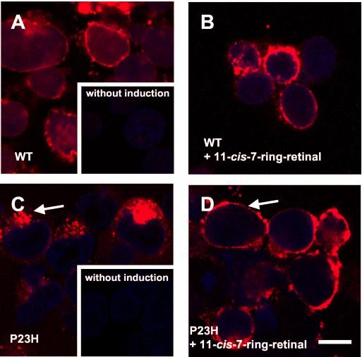
Fig. 7.

Subcellular localization of Rh and mutant P23H-opsin in HEK293 cells. Rh (red) was immunolabeled by anti-Rh monoclonal antibody 1D4. DNA (blue) was stained by Hoechst 33342. A, cells were fixed 24 h after inducing the expression of Rh. The most intense labeling (red) was observed on the surface of cells. Inset, the cells were cultured without inducing the expression of Rh. No staining (red) is observed. B, cells were treated with 11-cis-7-ring retinals and cultured for 24 h. Intense labeling (red) was observed on the surface of cells. C, cells were fixed 24 h after inducing the expression of P23H-opsin. Intense labeling (red) was observed in intracellular structure. Inset, the cells were cultured without inducing the expression of P23H-opsin. No staining (red) is observed. D, cells were treated with 11-cis-7-ring retinals and cultured for 24 h. Intense labeling (red) was observed on the surface of cells. The bar indicates 10 μm.
