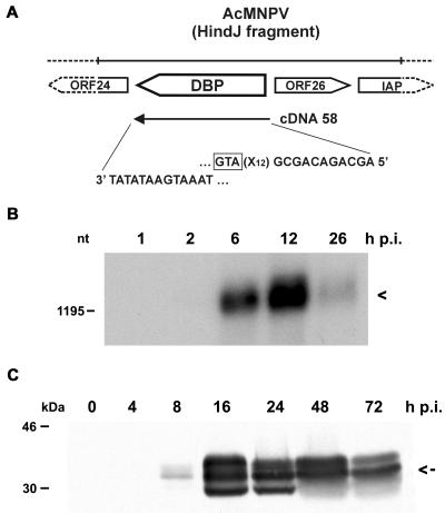
FIG. 3.
Characterization of DBP from AcMNPV. (A) Schematic representation of the dbp ORF on the AcMNPV genome. The sequences at the 5′ and 3′ ends of dbp cDNA clone 58 are indicated. IAP, inhibitor of apoptosis genes. The translational start codon of the DBP ORF (GTA) is boxed. (B) Polyadenylated RNA (10 μg) isolated from S. frugiperda cells at 1, 2, 6, 12, and 26 h p.i. was analyzed on a 1.2% agarose gel containing 2.2 M formaldehyde. The Northern blot was hybridized to DBP-specific cDNA clone 58. Arrowhead, transcript of about 1,300 nt. The position of the DNA size marker is given on the left. (C) Nuclear protein extracts were prepared from uninfected cells (lane 0) and from cells at 4, 8, 16, 24, 48, and 72 h p.i. Proteins were resolved on SDS-10% polyacrylamide gels and stained with anti-DBP antiserum. Arrow, middle-sized protein band of about 34 kDa. Protein size markers are given on the left.

