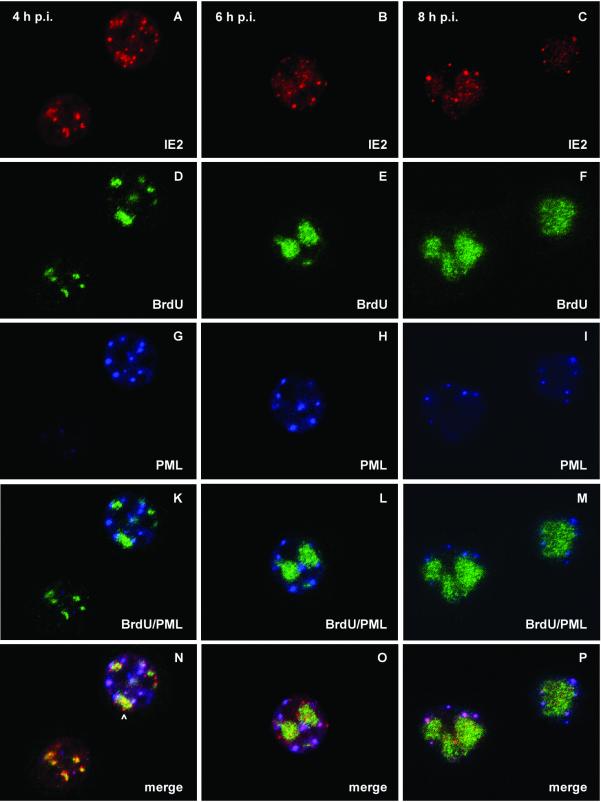
FIG. 5.
Localization of IE2, PML, and viral DNA replication sites after AcMNPV infection of TN-368 cells. TN-368 cells were infected with recombinant virus AcMNPV-PML/e. Cells were fixed at 4, 6, and 8 h p.i. and triple stained with rabbit anti-IE2 antiserum (red), rat BrdU MAb (green), and mouse MAb 5E10 (blue) to visualize PML. The arrowhead indicates IE2 dot clusters. Confocal images with double (K, L, and M) and triple merges (N, O, and P) are shown.

