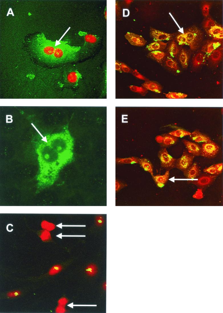
FIG. 12.
Vero cells were either infected with IBV (A) or transfected with pCi-IBV-N (B) and then incubated for 24 h; IBV proteins are shown in green, and nuclear DNA was visualized with PI (A). Cleavage furrows are indicated by an arrow in panels A and B. (C) Mock-transfected cells were stained for fibrillarin (green) and nuclear DNA (red) by using PI; dividing cells are indicated by an arrow. (D and E) Primary chicken kidney cells were infected with IBV for 8 h, and cells were stained for IBV (green) or actin (red). An arrow indicates a cleavage furrow in panel D and a nucleolus in E. Magnifications: A and B, ×60; C, D, and E, ×15.

