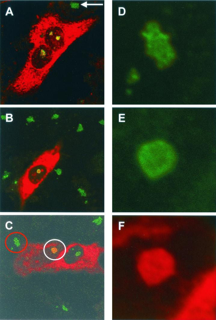
FIG. 3.
HeLa (A and C to F) or Vero (B) cells were transfected with either pCi-MHV-N (B to F) or pCi-IBV-N (A) and then fixed and analyzed by indirect immunofluorescence with rabbit anti-MHV or anti-IBV polyclonal sera (red). The nucleolus was detected with anti-fibrillarin (human) mouse monoclonal antibody (green). The structures identified by the red and white circles in panel C were resolved an additional 12 times (panel D and panels E and F, respectively). Colocalization when it occurs is shown in yellow. The arrow indicates a nucleolus in an adjacent untransfected cell. Magnifications: A, B, and C, ×60 (zoom, ×2).

