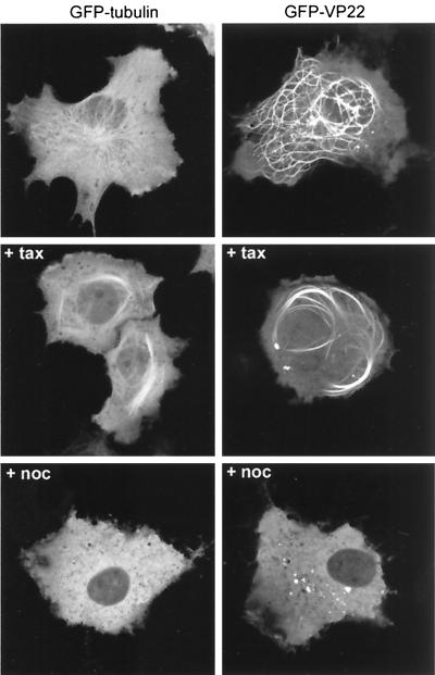
FIG. 1.
Comparison of GFP-tubulin and GFP-VP22 localization in live cells. COS-1 cells were transfected with expression vectors for either GFP-tubulin or GFP-VP22. Twenty hours later the cells were left untreated (top panels), incubated in medium containing taxol (+ tax), or incubated in medium containing nocodazole (+ noc). Live expressing cells were then imaged 2 h later by confocal microscopy.

