
Fig. 4.
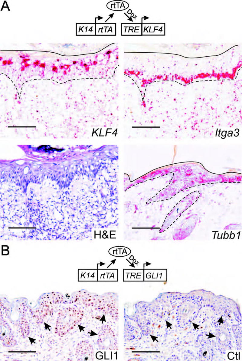
TRE-KLF4 and TRE-GLI1 transgene expression in hyperplastic skin. K14-rtTA;TRE-KLF4 (A) or K14-rtTA;TRE-GLI1 (B) bitransgenic mice were induced with dox, and cryosections of hyperplastic skin were analyzed. This 21 day induction was performed independently of the experiment shown in Fig. 3. A, mRNA in situ hybridization analysis using the indicated anti-sense probe. Positive staining is indicated as a red precipitate. Orientation lines correspond to the skin surface (solid lines) or to the DEJ (dashed lines). An H&E-stained cryosection is shown at bottom left. B, Immunostain of GLI1-induced hyperplastic lesions with GLI1 antibody. Normal rabbit immunoglobulin served as a control (Ctl). Staining by anti-GLI1 was low or negative on normal, interfollicular mouse skin (not shown). Arrows indicate the DEJ. Scale bars, 100μ.