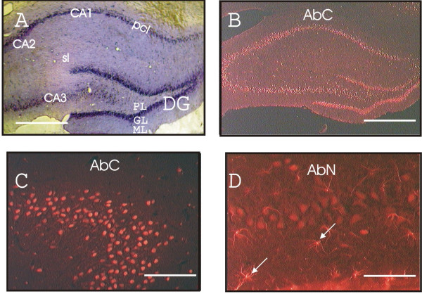
Figure 2.

Expression of ZAS3 in neuronal cell bodies and astrocytes at the hippocampal formation. (A) Rat coronal brain section (Bregma -3.80) stained with cresyl violet; (B) immunostaining of a similar brain sections with AbC; (C) CA2 region stained with AbC; and (D) CA3 region stained with AbN. Star-like AbN-positive cells are highlighted with arrows. pcl, pyramidal cell layer; sl, stratum lucidum; PL, GL and ML, polymorphic, granule and molecular cell layer, respectively, of the dentate gyrus. Scale bars represent 2 mm (A and B); 0.2 mm (C); 0.1 mm (D).
