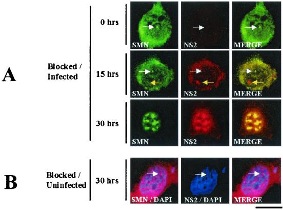
FIG. 5.
SMN and NS2 colocalize in SAABs at 30 h post-MVM infection. (A) SMN (primary, anti-SMN rabbit polyclonal sera; secondary, fluorescein isothiocyanate/green) and NS2 (primary: mouse monoclonal antibody; secondary: tetramethyl rhodamine isothiocyanate/red) are indicated. Synchronized infections were obtained by performing an isoleucine-aphidicolin double block on A92L cells prior to infection. Immunofluorescence experiments were performed on MVM-infected cells 0, 15, and 30 h postrelease (27). Nuclear Cajal bodies (white arrows) and NS2-positive/SMN-negative nuclear bodies (yellow arrows) are indicated. (B) SAAB formation is a consequence of viral infection. Smn localization was examined in synchronized, noninfected A92L cells 30 h postrelease. SMN (primary: anti-SMN rabbit polyclonal sera; secondary: tetramethyl rhodamine isothiocyanate/red) and NS2 (primary: mouse monoclonal antibody; secondary: fluorescein isothiocyanate/green) are indicated. Cell nuclei were counterstained with 4′,6′-diamidino-2-phenylindole (blue). The bar represents ≈30 μm.

