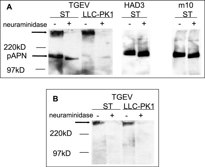
FIG. 2.
Binding of TGEV and the mutants HAD3 and m10 to cell surface proteins. Proteins were isolated from ST or LLC-PK1 cells by surface biotinylation and either mock treated (−) or neuraminidase treated (+). Following electrophoretic separation under nonreducing (A) or reducing (B) conditions, the proteins were transferred to nitrocellulose. The immobilized proteins were incubated with purified virus, and bound virus was detected by an enzyme-linked immunoassay. On the left side, the positions of molecular mass markers are indicated. Arrows point to aminopeptidase N and to the high-molecular-mass sialoglycoprotein recognized by TGEV.

