Abstract
Since the 1997 H5N1 influenza virus outbreak in humans and poultry in Hong Kong, the emergence of closely related viruses in poultry has raised concerns that additional zoonotic transmissions of influenza viruses from poultry to humans may occur. In May 2001, an avian H5N1 influenza A virus was isolated from duck meat that had been imported to South Korea from China. Phylogenetic analysis of the hemagglutinin (HA) gene of A/Duck/Anyang/AVL-1/01 showed that the virus clustered with the H5 Goose/Guandong/1/96 lineage and 1997 Hong Kong human isolates and possessed an HA cleavage site sequence identical to these isolates. Following intravenous or intranasal inoculation, this virus was highly pathogenic and replicated to high titers in chickens. The pathogenesis of DK/Anyang/AVL-1/01 virus in Pekin ducks was further characterized and compared with a recent H5N1 isolate, A/Chicken/Hong Kong/317.5/01, and an H5N1 1997 chicken isolate, A/Chicken/Hong Kong/220/97. Although no clinical signs of disease were observed in H5N1 virus-inoculated ducks, infectious virus could be detected in lung tissue, cloacal, and oropharyngeal swabs. The DK/Anyang/AVL-1/01 virus was unique among the H5N1 isolates in that infectious virus and viral antigen could also be detected in muscle and brain tissue of ducks. The pathogenesis of DK/Anyang/AVL-1/01 virus was characterized in BALB/c mice and compared with the other H5N1 isolates. All viruses replicated in mice, but in contrast to the highly lethal CK/HK/220/97 virus, DK/Anyang/AVL-1/01 and CK/HK/317.5/01 viruses remained localized to the respiratory tract. DK/Anyang/AVL-1/01 virus caused weight loss and resulted in 22 to 33% mortality, whereas CK/HK/317.5/01-infected mice exhibited no morbidity or mortality. The isolation of a highly pathogenic H5N1 influenza virus from poultry indicates that such viruses are still circulating in China and may present a risk for transmission of the virus to humans.
Wild waterfowl and shorebirds provide a reservoir for all 15 influenza A virus hemagglutinin (HA) subtypes, but infections in these species generally do not produce clinical signs (2, 3, 48, 54). From this reservoir, a few influenza viruses from the H5 and H7 subtypes continue to emerge in domestic poultry, and selective pressure of these viruses to adapt to a new host can result in the emergence of a virulent virus (46). Such viruses cause a disease formerly known as fowl plague but now more commonly referred to as highly pathogenic avian influenza (HPAI) (4, 50). In poultry, HPAI viruses cause multiorgan systemic disease resulting in high morbidity and mortality in chickens. In 1997 in Hong Kong, HPAI (H5N1) viruses circulated in poultry farms and later in wholesale and retail poultry markets. The HA gene of the Hong Kong/97 H5N1 viruses was closely related to that of the A/goose/Guangdong/1/96 (H5N1) virus and contained multiple basic amino acids adjacent to the cleavage site between HA1 and HA2 (44, 47). This molecular characteristic of influenza virus has been identified as a major virulence factor for chickens and turkeys by giving the virus the ability to replicate in a wide range of cell types, resulting in severe disseminated disease and high mortality (36, 39). Further characterization of the HPAI H5N1 viruses showed that all eight gene segments are closely related to those of the pathogenic H5N1 influenza viruses that affected humans later in 1997 (6, 31, 44, 47). Six deaths were recorded from 18 confirmed hospitalized cases. This was the first report of a purely avian influenza virus (AIV) causing respiratory disease and death in humans (9, 10, 12, 13, 47, 57). Subsequent epidemiologic investigations revealed no evidence of effective human-to-human transmission; the human cases were apparently the result of multiple independent transmissions of the H5N1 viruses from infected poultry to humans (7, 23, 33). Therefore, chickens could serve as an amplifying intermediate host for the transmission of AIVs from wild aquatic birds to humans.
Although no H5N1 viruses have been isolated from humans since December 1997, genetically related viruses are still being detected in domestic poultry in Hong Kong and southern China. In March 1999, four H5N1 viral isolates were obtained by swabbing cages that housed geese in Hong Kong. These isolates, designated A/Environmental/Hong Kong/437/99, contained HAs that are closely related to the 1997 human/poultry H5N1 viruses and the Goose/Guangdong/1/96 virus (8). In addition to H5N1 viruses, the H9N2 isolate, A/quail/Hong Kong/G1/97, possessing six internal genes that are closely related to the 1997 human/poultry H5N1 viruses, is currently widespread in domestic poultry in Hong Kong and southern China (16, 17). The isolation of H9N2 viruses from five humans with respiratory illnesses in southern China and two pediatric cases in Hong Kong in 1999 raised concern of another AIV that could cross the species barrier to replicate in humans (16, 18, 19, 27, 35). More recently, H5N1 isolates containing Goose/Guangdong/1/96-like HA and NA genes have been detected in several retail live-bird markets in the Hong Kong Special Administrative Region (SAR). This has resulted in a second depopulation of poultry in 4 years in an attempt to remove the source of infection (World Health Organization, Disease outbreaks reported, 18 May 2001).
The avian Hong Kong H5N1 and H9N2 viruses were unique in their ability to replicate in humans without prior adaptation in a mammalian host. The BALB/c mouse has been used as a mammalian model system to study H5N1 and H9N2 virus pathogenesis (14, 29, 30). These viruses replicate efficiently in the lungs of mice, but clear differences in pathogenicity were observed among the virus isolates (14, 15, 19, 29, 30). As for the H5N1 Hong Kong/97 viruses, all 17 isolates were highly pathogenic in chickens; however, not all were lethal in mice. Some of the H5N1 viruses replicated only in the respiratory tract, whereas others were lethal in mice and infectious virus was detected in multiple organs (24, 25, 29). Taken together, this suggests that the HA cleavage site is not the primary genetic determinant associated with high pathogenicity of H5N1 influenza viruses in mice.
The introduction of H5N1 AIVs to humans in Hong Kong in 1997 and the continued presence of H5N1-like viruses in southern China emphasize the importance of continued surveillance, isolation, and characterization of virus subtypes and variants present in poultry. This paper reports the recovery of an HPAI H5N1 influenza virus from domestic duck meat. We provide molecular characterization and pathogenicity of A/Duck/Anyang/AVL-1/01 virus and compare it with that of other H5N1 viruses isolated in Hong Kong since 1997. Genetic analysis showed that the HA gene is closely related to the H5N1 viruses that caused human infections in 1997. Like the 1997 H5N1 viruses, this virus was highly pathogenic in chickens and replicated efficiently in the lungs of mice without prior adaptation. Isolation of DK/Anyang/AVL-1/01 virus from muscle and brain tissue of experimentally infected ducks helps define the host range of this virus. To our knowledge, this is the first demonstration of an HPAI H5N1 influenza virus isolated from domestic duck meat and raises important public health implications.
MATERIALS AND METHODS
Viruses, isolation, and identification.
In South Korea, all imported poultry meat from China has been quarantined since HPAI virus was isolated in Hong Kong in May 2001. During this quarantine, an influenza A virus was isolated from imported Cherry Valley Pekin duck (Anas platyrhynchos) meat. Duck meat processed at a food factory in Shanghai, mainland China, was brought from farms located in the Shanghai region. Complying with procedures of national regulation, five samples of meat (200 to 500 g) were taken from each shipment container of 7,500 duck carcasses. Random sampling was performed on breast, wing, thigh, and back of whole duck carcasses. Samples of frozen duck meat containing the skin were kept in a sterile plastic bag and maintained frozen at −70°C. Each sample was frozen and thawed three times and the extracted fluid was collected and clarified by low-speed centrifugation. The supernatant was filtered through a sterile membrane filter with a 0.45-μm pore size, and a 0.2-ml volume of the filtrate was inoculated into the allantoic cavity of each of five 10-day-old embryonated specific-pathogen-free (SPF) hens' eggs. Inoculated eggs were incubated at 37°C for 30 h. After that, allantoic fluids from dead eggs were harvested and tested for hemagglutination activity. Seven shipment containers out of 15 containers imported from a Shanghai food factory were positive for influenza virus, yielding seven isolates. All seven isolates were typed as an influenza A H5N1 virus by means of hemagglutination inhibition and neuramindase inhibition tests with a panel of antisera (provided by OIE Reference Laboratory, Veterinary Laboratory Agency, Surrey, United Kingdom). The sequence homology at the hemagglutinin cleave site (sequenced 290 bp) of the seven isolates was 99.6 to 100%, indicating a similar origin. The isolate was designated as A/Duck/Anyang/AVL-1/01 (DK/Anyang/AVL-1/01). Laboratory contamination on isolation by the National Veterinary Research and Quarantine Service, Anyang, Korea, was not likely since no other virology work with HPAI was conducted at this laboratory. Additional influenza H5N1 viruses used at Southeast Poultry Research Laboratory in this study were A/Environment/Hong Kong/437/99 (Env/HK/437-6/99), A/Chicken/Hong Kong/317.5/01 (CK/HK/317.5/01), and A/Chicken/Hong Kong/220/97 (CK/HK/220/97) (all received courtesy of Les Sims, Agriculture and Fisheries Department, Hong Kong). Virus stocks were propagated 24 to 30 h in the allantoic cavity of eggs at 37°C. Infectious allantoic fluid was aliquoted and stored at −70°C. Serial titration was performed in eggs and incubated at 37°C for 24 to 30 h. After that, allantoic fluids from eggs were harvested and 50% egg infectious dose (EID50) titers were determined by testing hemagglutination activity. Titration endpoints were calculated by the method of Reed and Muench (38). All four H5N1 viruses had high infectivity titers in eggs (107.9 to 109.0 log10 EID50/ml). All experiments using infectious HPAI H5N1 viruses, including work with animals, were conducted using biosafety level 3+ (BSL-3+) Ag containment procedures (5). All personnel were required to wear a powered air protection respirator with HEPA-filtered air supply (RACAL Health and Safety Inc., Frederick, Md.).
Molecular cloning and sequencing of influenza virus genes.
RNA from the isolate sequenced in this study was extracted with Trizol LS reagent (Life Technologies, Rockville, Md.) from infectious egg allantoic fluid prior to reverse transcriptase PCR (RT-PCR) amplification. The RT-PCR amplification was performed with the Onestep RT-PCR kit (Qiagen, Valencia, Calif.) with incubation steps of 50°C for 30 min and 95°C for 15 min and then 30 cycles of annealing at 51 to 56°C for 15 s, extension at 72°C for 60 s, and denaturation at 94°C for 30 s. The NS, M, and NP gene segments were amplified with primers to the conserved 12 and 13 bp present on the 5′ and 3′ end of each viral segment. The HA, NA, PB1, PB2, and PA genes were RT-PCR amplified with specific primers also from the noncoding sequence of each gene segment. For all eight viral genes, the full coding sequence was amplified. The PCR product was electrophoresed in an agarose gel, and the DNA corresponding in size to the gene segment of interest was extracted with the Agarose Gel DNA extraction kit (Roche, Indianapolis, Ind.). The primers used included 12-bp 5′ extensions that allowed the PCR product to be cloned with the ligation-independent cloning system pAmp1 (Life Technologies). Colonies were screened by PCR with internal primers. Positive cultures were grown overnight and plasmids were extracted using the Qiaprep spin miniprep kit (Qiagen). Plasmids were sequenced using the ABI PRISM Bigdye terminator sequencing kit (Perkin-Elmer, Foster City, Calif.) run on an ABI 3700 automated sequencer (Perkin-Elmer). Since only a single clone for each gene was included in the sequence analysis, the potential for Taq polymerase-introduced sequence errors was possible. However, because of the quasispecies nature of influenza virus, it is difficult to determine a most correct sequence for any given isolate.
The sequencing information was compiled with the Seqman program (DNASTAR, Madison, Wis.), and the nucleotide sequences were compared initially with Megalign program (DNASTAR) using the Clustal alignment algorithm. Pairwise sequence alignments were also performed in the Megalign program to determine sequence similarity between DK/Anyang/AVL-1/01 and other published sequences for each gene segment. Phylogenetic comparisons of the aligned sequence for each gene segment were generated using the maximum parsimony method, with 100 bootstrap replicates in a heuristic search using the PAUP 4.0b4 software (Sinauer Associates, Inc, Sunderland, Mass.).
Chicken experiments.
Four-week-old SPF white Plymouth Rock (WPR) chickens were used in pathogenicity studies using established procedures (34, 52). The chickens were housed in stainless steel isolation cabinets that were ventilated under negative pressure with HEPA-filtered air, and care was provided as required by the Institutional Animal Care and Use Committee based on the Guide for the Care and Use of Agricultural Animals in Agricultural Research and Teaching. Feed and water were provided ad libitum. Eight chickens were inoculated by the intravenous (i.v.) route with 0.2 ml of a 1:10 dilution of a bacteria-free, allantoic fluid containing 108.0 EID50 of DK/Anyang/AVL-1/01 virus. The pathogenicity test also included 11 chickens inoculated intranasally (i.n.) with 106.0 EID50 of DK/Anyang/AVL-1/01 virus. For the latter group of birds, three chickens were euthanatized on day 2 postinoculation (p.i.) with sodium pentobarbital (100 mg/kg of body weight) given i.v. These birds, along with three additional birds that died from infection on day 3 p.i., were evaluated for gross lesions and tissues were collected for virus isolation, histopathology, and immunohistochemistry (IHC). Procedures for histopathology and IHC followed those previously described (37, 49). Briefly, lungs, bursa, kidneys, adrenal gland, thymus, thyroid, brain, liver, heart, pancreas, intestine, spleen, trachea, thigh, and breast tissue were collected from a total of six DK/Anyang/AVL-1/01-infected chickens. Tissues were fixed in 10% neutral buffered formalin solution, sectioned, and stained with hematoxylin-and-eosin. Duplicate sections were stained by IHC methods to determine influenza viral antigen distribution in individual tissues. A monoclonal antibody against influenza A virus nucleoprotein (P13C11), developed at Southeast Poultry Research Laboratories, was used as the primary antibody in a streptavidin-biotin-alkaline-phosphatase-complex IHC method as previously described (37, 49). Portions of the brain, lung, kidney, thigh, and breast tissue were stored frozen at −70°C and titers of infectious virus were subsequently determined as previously described (8). Briefly, tissues were weighed and homogenized in brain heart infusion (BHI) medium and clarified homogenates were titrated for virus infectivity in eggs.
Duck experiments.
Two-week-old Pekin white ducks (A. platyrhynchos) (Privett hatchery, Portales, N.Mex.) (eight per group) were inoculated i.n. with 106.0 EID50 of each of the respective influenza viruses administered in a volume of 0.1 ml. In addition, four control ducks were inoculated with 0.1 ml of sterile allantoic fluid diluted 1:300 in BHI medium and served as the mock-infected controls. Ducks were observed daily for clinical signs of disease. Oropharyngeal and cloacal swabs were collected from four ducks each day from 1 to 7 days p.i. and from two ducks on days 10 and 14 p.i. Oropharyngeal and cloacal swabs and tissues were collected at 4 and 13 days p.i. from four control ducks. Two ducks were euthanatized and necropsied at 2, 4, 7, and 14 days p.i. Gross lesions were recorded, and tissues (brain, lung, kidney, and skeletal muscle) were collected separately from each duck for virus isolation, histopathology and IHC as described above. Skeletal muscle from the proximal shank (pars interna of the gastrocnemius muscle), larynx, and/or periorbital region was collected for histopathology and for the presence of viral antigen. For virus isolation, skeletal muscle from the proximal shank was stored at −70°C and subsequently processed as described above.
Mouse experiments.
Male BALB/c mice 6 to 8 weeks old (Simonsen Laboratories, Gilroy, Calif.) were anesthetized with ketamine-xylazine (1.98 and 0.198 mg per mouse, respectively). In the first experiment, 12 mice per group were inoculated i.n. (50 μl) with 106.0 EID50 of DK/Anyang/AVL-1/01, CK/HK/220/97, or CK/HK/317.5/01 (H5N1) influenza virus stocks diluted in phosphate-buffered saline (PBS). An additional group of mice received diluent PBS in place of virus and served as the mock-infected controls. Nine mice per group were monitored daily for morbidity (measured by weight loss) and death for 14 days p.i. Blood samples (20 to 40 μl) were collected from infected mice on days 0, 3, 5, 7, and 9 p.i. Absolute leukocyte counts were determined with a hemocytometer on heparinized blood diluted 1:10 with Turks solution (2% acetic acid, 0.01% methylene blue). Cell numbers were determined in triplicate from two individual mice. For differential counts, peripheral blood was obtained from two or three mice on the days indicated. Two blood smears from each mouse were stained with Hema-3 stain (Fisher Diagnostics, Orangeburg, N.Y.), and the numbers of monocytes, polymorphonuclear neutrophils, and lymphocytes were determined. At least 100 cells were counted for each slide at a magnification of ×1,000. Three mice from each group were euthanatized on day 4 p.i. and evaluated for gross lesions. The sinuses, bone marrow, brain, testes, thymus, kidneys, adrenal gland, lungs, vesicular gland, muscle, heart, liver, spleen, pancreas, intestine, and stomach were collected for histopathology and IHC, as described above.
In a second experiment, 12 mice per group were inoculated with the same dose of DK/Anyang/AVL-1/01, CK/HK/220/97, or CK/HK/317.5/01 as described in the first experiment. Four days later, four mice were euthanatized and whole lungs, kidneys, brains, and tracheas (5 mm in length) were collected and homogenized in 1 ml of cold PBS. The solid debris was removed by brief centrifugation before homogenates were titrated for virus infectivity in eggs from initial dilutions of 1:10 (lung and trachea) or 1:2 (kidney and brain). The limit of virus detection was 101.2 EID50/ml for lung and trachea and 100.8 EID50/ml for other tissues. The remaining nine mice per group were monitored daily for morbidity (measured by weight loss) and death for 14 days p.i.
Nucleotide sequence accession numbers.
Sequence data were submitted to GenBank with accession numbers AF468837 to AF468844 and AY075027 to AY075036.
RESULTS
Phylogenetic analysis of DK/Anyang/AVL-1/01.
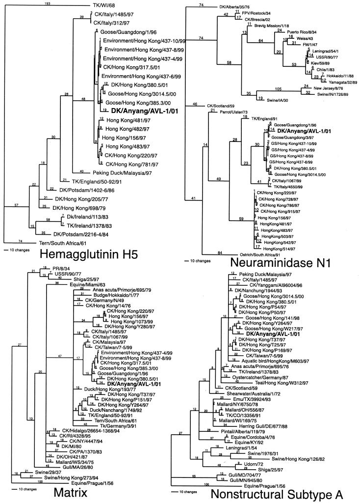
Phylogenetic sequence analysis of the eight gene segments showed that the DK/Anyang/AVL-1/01 isolate has a unique assortment of viral gene segments. The hemagglutinin gene clustered with the H5 Goose/Guangdong/1/96 lineage and the Hong Kong/97 chicken and human isolates (Fig. 1). The hemagglutinin gene had the highest sequence similarity with the virus isolate Env/HK/437-6/99 (Table 1), which was associated with geese entering Hong Kong for slaughter in March 1999 (8). The sequence at the hemagglutinin cleavage site for DK/Anyang/AVL-1/01 was identical to the other HPAI viruses with the insertion of four basic amino acids (aa) in addition to three other basic amino acids.
FIG. 1.
Phylogenetic trees of the nucleotide sequences of HA subtype 5, neuraminidase subtype 1, matrix, and nonstructural subtype (group) A, including the isolate DK/Anyang/AVL-1/01 and representative human, swine, equine, and avian influenza virus gene sequences when appropriate. The trees were generated with the PAUP 4 computer program with bootstrap replication (100 bootstraps) and a heuristic search method. The HA tree is rooted to TK/WI/68, the neuraminidase tree is rooted to DK/Alberta/35/76, and the matrix and nonstructural trees are rooted to Equine/Prague/1/56. Branch lengths are included on the tree. Standard two-letter postal codes are used for states in the United States. Abbreviations: TK, turkey; CK, chicken; DK, duck. For isolates without a species, it is assumed to be an isolate from a human.
TABLE 1.
Sequence similarity between gene segments of DK/Anyang/AVL-1/01 and other influenza isolates
| Gene product | nta similarity (%) | Isolate | aa similarity (%) | Isolate |
|---|---|---|---|---|
| Hemagglutinin H5 | 98.4 | Env/Hong Kong/437-6/99 | 98.4 | DK/Hong Kong/380.5/01 |
| Neuraminidase N1 | 98.0 | Goose/Guangdong/1/96 | 98.4 | Goose/Guangdong/3/97 |
| PB1 | 95.0 | CK/Taiwan/7-5/99 | 98.9 | Gull/MD/704/77 |
| PB2 | 94.6 | Env/Hong Kong/437-6/99 | 98.5 | Seal/MA/133/82 |
| PA | 93.8 | Swine/Hong Kong/81/78 | 98.6 | TK/MN/833/78 |
| NP | 97.4 | Env/Hong Kong/437-8/99 | 99.0 | Env/Hong Kong/437-8/99 |
| MA, M1 | 97.0 | Goose/Guangdong/1/96 | 98.0 | Goose/Guangdong/1/96 |
| NS, NSI | 96.9 | DK/Nanchang/1944/93 | 96.1 | Anas acuta/Primorje/695/76 |
nt, nucleotide.
Phylogenetic analysis of the neuraminidase gene also showed that DK/Anyang/AVL-1/01 clustered with the Goose/Guangdong/1/96 virus lineage (Fig. 1). The Goose/Guangdong/1/96 virus lineage includes the Env/HK/437-6/99, and the recent 2000 to 2001 H5N1 viruses from Hong Kong, Goose/Hong Kong/3014.5/00, CK/HK/317.5/01, and DK/Hong Kong/380.5/01. The highest sequence homology was to Goose/Guangdong/1/96 (Table 1) and not to later viruses from this lineage. However, the DK/Anyang/AVL-1/01 isolate had a 20-aa deletion in the stalk region that was not found in the other viruses. The DK/Anyang/AVL-1/01 neuraminidase gene was distinctly different from the H5N1 Hong Kong/97 chicken and human isolates, although these viruses also had a 19-aa stalk deletion in the same region of the neuraminidase protein. For the internal genes, the matrix (Fig. 1) and nucleoprotein genes did cluster loosely with the Goose/Guangdong/1/96 lineage of viruses, although the highest nucleotide sequence similarity was only 97 and 97.4%, respectively, with this group of viruses (Table 1). The nonstructural genes (Fig. 1) and all three polymerase genes did not cluster closely with any other influenza viruses (Table 1), although all four genes were in the avian Eurasian lineage of influenza viruses.
Pathogenicity of DK/Anyang/AVL-1/01 in chickens.
We determined the pathogenicity of DK/Anyang/AVL-1/01 virus in 4-week-old WPR chickens. By criteria, AIVs that kill 75% or more of eight i.v. inoculated chickens within 10 days are classified as highly pathogenic (34, 52). In addition to the i.v. test, chickens were inoculated i.n. with 106.0 EID50 of DK/Anyang/AVL-1/01 virus. Intravenous or i.n. inoculation with DK/Anyang/AVL-1/01 virus caused 100% (eight of eight) mortality, and disease signs observed were typical of those seen in chickens infected with H5N1 Hong Kong/97 viruses (44). The mean death times were 3 days following i.v. inoculation and 2.9 days following i.n. inoculation. Tissues from six chickens that died or were euthanatized on days 2 and 3 days p.i. following i.n. inoculation were evaluated for gross lesions, histopathology, infectious virus, and viral antigen expression. Titration of tissues revealed high titers (6.2 to 6.7 log10 EID50/g of tissue) of infectious virus from brain, lung, kidney, and thigh tissue, with lower titers in the breast tissue (5.3 to 5.5 log10 EID50/g). On days 1 to 3 p.i., relatively high titers of infectious virus could also be isolated from oropharyngeal swabs (4.4 to 4.8 log10 EID50/ml), whereas DK/Anyang/AVL-1/01 virus shedding from the cloaca (1.1 to 3.1 log10 EID50/ml) was considerably less.
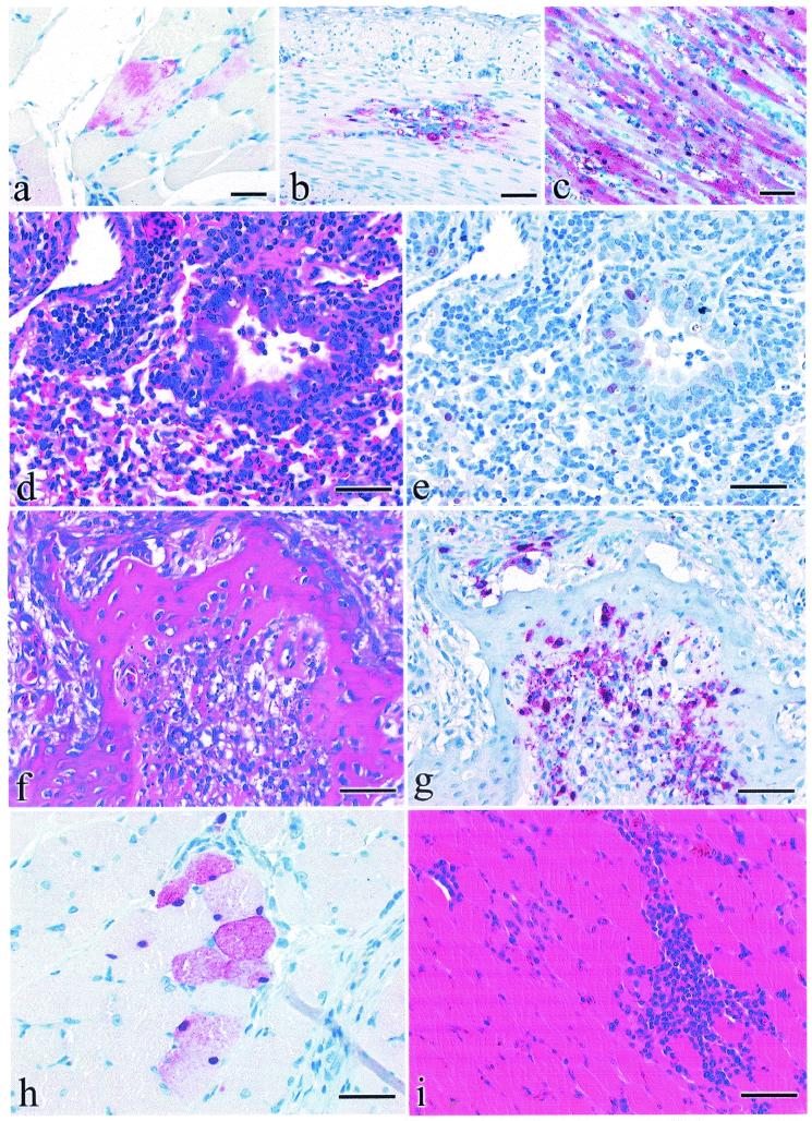
As with the Hong Kong H5N1 viruses, DK/Anyang/AVL-1/01 virus produced similar systemic lesions in chickens following i.n. inoculation. The lesion severity and viral antigen quantity were less in birds euthanatized than in those that died on days 2 and 3 p.i., but lesion distribution was similar. Of interest was skeletal muscle, where birds that died had multifocal moderately severe degeneration and necrosis of myofibers with commonly associated influenza viral antigen in nuclei and cytoplasm (Fig. 2a ). The euthanatized birds lacked lesions in skeletal muscle and viral antigen was sporadic to rare in myofibers (Fig. 2b). Interestingly, birds that died had influenza viral antigen in smooth muscle cells of the small and large intestines and abundant viral antigen in 25 to 75% of cardiac myocytes (Fig. 2c). Other lesions identified included mild neuronal necrosis in the brain, mild-to-moderate histiocytic interstitial pneumonia with edema, mild-to-severe necrosis in the pancreas, mild-to-severe necrosis in adrenal glands, and mild-to-moderate nephrosis. Influenza viral antigen was identified in neurons, microglia, and ependyma of the brain; histiocytes and heterophils in the lungs; pancreatic acinar epithelium; adrenal corticotrophic cells; and kidney tubular epithelial cells. In addition, sporadic to abundant antigen was demonstrated in vascular endothelium throughout most visceral organs.
FIG. 2.
Experimental studies of chickens, ducks, and mice inoculated with DK/Anyang/AVL-1/01. Photomicrographs of hematoxylin-and-eosin-stained tissue sections (d, f, and i) or sections stained by IHC methods to demonstrate AIV (a to c, e, g, and h). (a) AIV antigen in cytoplasm of skeletal muscle fibers from a 4-week-old chicken that died 3 days after i.n. inoculation. Bar = 18 μm. (b) AIV antigen in cytoplasm and nuclei of smooth muscle fibers within the tunica muscularis of duodenum from a 4-week-old chicken that died 3 days after i.n. inoculation. Bar = 40 μm. (c) AIV antigen in cytoplasm and nuclei of cardiac muscle fibers from a 4-week-old chicken that died 2 days after i.n. inoculation. Bar = 40 μm. (f) Focal acute periosteal necrosis in pneumatic bone of the cranium in a 2-week-old duck euthanatized 2 days after i.n. inoculation. Bar = 35 μm. (g) AIV antigen in periosteal mesenchymal cells in a 2-week-old duck euthanatized 2 days after i.n. inoculation. Bar = 35 μm. (h) AIV antigen in perilaryngeal skeletal myocytes in a 2-week-old duck euthanatized 2 days after i.n inoculation with DK/Anyang/AVL-1/01 virus. Bar = 15 μm. (i) Focal myofiber degeneration with corresponding lymphohistiocytic myositis in perilaryngeal skeletal muscle in a 2-week-old duck euthanatized 7 days after i.n. inoculation. Bar = 35 μm. (d) Necrotizing bronchitis with neutrophilic inflammation and associated lymphohistiocytic alveolitis in a 4-week-old BALB/c mouse that was euthanatized 4 days after i.n. inoculation. Bar = 35 μm. (e) AIV antigen in nuclei of bronchial epithelium and type II pneumocytes in 4-week-old BALB/c mice that was euthanatized 4 days after i.n. inoculation. Bar = 35 μm.
Pathogenicity of Dk/Anyang/AVL-1/01 virus in Pekin white ducks.
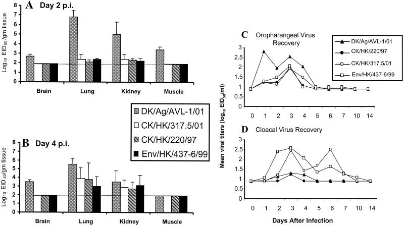
Since ducks were the source of the Dk/Anyang/AVL-1/01 virus, we determined the pathogenicity in this species and examined whether skeletal muscle could support virus replication. In addition, three highly pathogenic H5N1 isolates collected in 1997 (CK/HK/220/97), 1999 (Env/HK/437-6/99), and 2001 (CK/HK/317.5/01) were used to inoculate Pekin white ducks. In contrast to chickens, neither morbidity, mortality, nor gross lesions were observed in the ducks inoculated i.n. with any of the H5N1 isolates. We examined the kinetics of virus replication on days 2, 4, 7, and 14 p.i in brain, lung, kidney, and muscle tissue following an i.n. infection with the H5N1 isolates. As shown in Figure 3A and B, infection of ducks with each of the H5N1 viruses resulted in detectable virus in the lungs and kidneys on days 2 and 4 p.i. At 2 days p.i., infectious virus in the lungs and kidneys of ducks infected with the Dk/Anyang/AVL-1/01 virus was substantially higher than that in ducks infected with the other H5 viruses. None of the H5N1 viruses could be detected in these tissues on days 7 and 14 p.i. Interestingly, virus was also detected in brain and muscle tissue of Dk/Anyang/AVL-1/01-infected ducks but could not be detected with the other H5N1 viruses tested (Fig. 3A and B). Virus was recovered from the cloaca and oropharynx from each of the four H5N1 virus-infected groups. Figure 3C and D shows the mean virus titers from four birds that were determined on various days p.i. In general, each H5N1 virus isolate could be recovered from the cloaca and oropharynx during the first week of infection, although very little infectious virus could be recovered from cloacal swabs of CK/HK/220/97-infected and DK/Anyang/AVL-1/01-infected ducks (Fig. 3D). Higher levels of DK/Anyang/AVL-1/01 virus could be recovered from the oropharynx in comparison to the cloaca up to 4 days p.i. (Fig. 3C).
FIG. 3.
Comparison of mean titers of influenza A virus recovered from tissues. Ducks were infected with 106 EID50 of each virus; tissues were collected on days 2 (A) and 4 (B) p.i. and titers in eggs were determined. All isolation attempts without recovery of virus were given a value of 101.9 EID50. This represents the limit of virus detection (horizontal dotted line) for tissues. Mean log10 titers expressed as EID50/milliliter from oropharyngeal (C) and cloacal (D) swabs were sampled from four individual Pekin ducks on the days indicated. For swabs, the limit of virus detection was 100.9 EID50/ml
Histologic lesions observed in the ducks inoculated with any of the four viruses tended to be only mild to moderate; however, the broadest distribution of lesions and viral antigen was observed in the DK/Anyang/AVL-1/01-inoculated ducks. At 2 and 4 days p.i., ducks inoculated with DK/Anyang/AVL-1/01 virus consistently had mild to moderate multifocal necrotizing to heterophilic rhinitis, sinusitis, laryngitis, and tracheitis. Viral antigen was infrequently demonstrated in epithelial cells and submucosal leukocytes that corresponded to the foci of necrosis and inflammation in each of these organs. In addition, discrete foci of necrosis were observed in the pneumatic bone of the skull, and corresponding viral antigen was demonstrated in periosseous mesenchymal cells (Fig. 2f and g). Mild to moderate multifocal interstitial pneumonia consisting of mixed mononuclear infiltrates with fewer heterophils centered on the parabronchi was observed in the lungs of the ducks at both 2 and 4 days p.i. Viral antigen was demonstrated only in epithelial cells lining a bronchus and parabronchus of one duck on day 2 p.i. Histological lesions at 7 and 14 days p.i. in the respiratory tissues consisted of a similar distribution of mild to moderate submucosal lymphoplasmacytic infiltrates. Viral antigen was not demonstrated in any of the respiratory tissues collected at 7 and 14 days p.i.
Skeletal muscle from the proximal shank, larynx, and/or periorbital region was examined for lesions and for the presence of viral antigen. On days 2 and 4 p.i., hyalinization to necrosis of a few scattered individual to small clusters of skeletal myofibers was observed in all three samples of skeletal muscle. The distribution of viral antigen in the tissues was consistent with the distribution of the affected myofibers (Fig. 2h). Despite a lack of viral antigen, mild multifocal mononuclear infiltrates were observed in the skeletal muscle collected at 7 and 14 days p.i. (Fig. 2i). However, contrary to the chickens, viral antigen was not observed in smooth myocytes in the DK/Anyang/AVL-1/01-inoculated ducks at any time. Mild lesions also were observed in the brain of both ducks sampled on day 4 p.i. and one duck sampled on day 7 p.i. These lesions included mild perivascular lymphoplasmacytic cuffs around a few localized cerebral vessels and associated small foci of gliosis. Viral antigen was demonstrated only in the affected ependymal cells of a single duck sampled at 4 days p.i.
In the present study, lesions in the ducks inoculated with the CK/HK/220/97 virus resembled those previously described for 4-week-old Pekin ducks (37). These lesions were confined to the respiratory tract and consisted of mild epithelial necrosis to lymphoplasmacytic inflammation of the air sac, larynx, trachea, and nasal cavity and a mild to moderate interstitial pneumonia, which was observed in one duck on day 4 p.i. and two ducks on day 7 p.i. Viral antigen was demonstrated in epithelial cells lining the nasal cavity and trachea of one duck sampled on day 4 p.i. Similarly, lesions produced by the CK/HK/317.5/01 virus or the Env/HK/437-6/99 virus were of similar distribution and severity as those produced by the CK/HK/220/97 virus.
Pathogenicity of DK/Anyang/AVL-1/01 virus in mice.
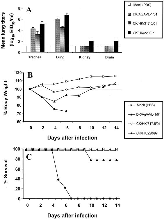
We previously demonstrated that the H5N1 Hong Kong/97 chicken and human isolates differed from other HPAI H5 viruses in their high pathogenicity for mice (14). To determine the pathogenicity of DK/Anyang/AVL-1/01 and CK/HK/317.5/01 viruses in a mammalian host, BALB/c mice were inoculated i.n. and virus replication, morbidity (measured by weight loss), and mortality were determined. For comparison, a group of mice were infected with the highly pathogenic CK/HK/220/97 virus, previously shown to cause 100% lethality in mice (14). Infection of mice with each of the H5N1 viruses resulted in high titers of virus in the trachea and lungs on day 4 p.i. (Fig. 4A). Virus was present in extrapulmonary tissues, including the brain and kidneys of mice infected with the highly pathogenic CK/HK/220/97 virus; however, mice infected with DK/Anyang/AVL-1/01 or CK/HK/317.5/01 virus had undetectable titers (≤100.8 EID50/ml) in these tissues. CK/HK/220-infected mice and, to a lesser extent, DK/Anyang/AVL-1/01-infected mice showed signs of illness, such as ruffled fur and hunched posture, and began to lose weight 2 days after infection (Fig. 4B). Weight loss continued rapidly in CK/HK/220-infected mice and mortality reached 100% by day 7 (Fig. 4C). With the exception of two of nine (22%) mice that succumbed to infection, the majority of the DK/Anyang/AVL-1/01-infected mice began to recover after day 4 p.i. All CK/HK/317.5/01-infected mice survived the infection and displayed only slight weight reduction on day 4 p.i. In a second mouse experiment, DK/Anyang/AVL-1/01 virus induced 33% (three of nine) mortality, whereas CK/HK/317.5/01 virus induced no (zero of nine) mortality.
FIG. 4.
Comparison of lung virus titers (A), weight loss (B), and lethality (C) of BALB/c mice infected with 106.0 EID50 of DK/Anyang/AVL-1/01, CK/HK/317.5/01, or CK/HK/220/97 or mock infected. Four to five mice from each virus-infected group were euthanatized on day 4 p.i., and titers in individual lung, trachea, kidney, and brain tissue in embryonated chicken eggs were determined. The limit of virus detection was 101.2 EID50/ml (dotted line). The remaining seven mice from each group were observed for weight loss and mortality through a 14-day observation period.
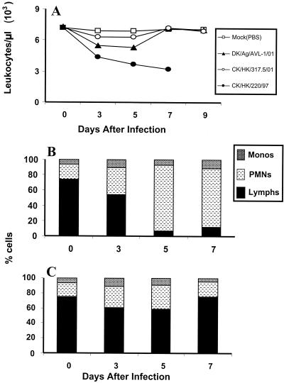
We next evaluated whether infection with DK/Anyang/AVL-1/01 resulted in alteration in peripheral blood leukocyte counts. Mice were bled on alternate days during the first week of infection and the total number of leukocytes and differential blood counts were determined. Leukopenia in CK/HK/220-infected mice was detected from day 3 p.i. and was statistically significant on day 7 p.i. relative to the leukocyte counts of mice from the DK/Anyang/AVL-1/01, CK/HK/317.5/01, or mock-infected groups (Fig. 5A). Furthermore, differential blood counts revealed that lymphocyte numbers in CK/HK/220-infected mice dropped up to 92% by day 5 p.i. and remained low until the death of these mice (Fig. 5B). In contrast, DK/Anyang/AVL-1/01-infected mice displayed a transient drop (10 to 22%) in lymphocyte numbers on days 3 and 5 p.i., with recovery to normal levels by day 7 (Fig. 5C).
FIG. 5.
Kinetic analysis of circulating leukocytes (A) and blood differential counts (B and C) following H5N1 infection. Two to three mice were infected i.n. with 106.0 EID50 of DK/Anyang/AVL-1/01 (▴), CK/HK/317.5/01 (○), or CK/HK/220/97 (•) or were mock infected (□). Total white blood cell counts were determined by microscopic counting of leukocytes in heparinized whole blood samples diluted with Turks solution. Blood smears were stained with Hema-3 differential stain, and the percentages of monocytes, polymorphonuclear neutrophils (PMNs), and lymphocytes in CK/HK/220/97-infected (B) and DK/Anyang/AVL-1/01-infected (C) mice were determined.
Three mice from each group were euthanatized 4 days after inoculation and evaluated for gross lesions, histopathologic changes, and viral antigen expression. Mice infected with DK/Anyang/AVL-1/01 had a slight reduction in visceral body fat content and gross pneumonic lesions in 20 to 33% of the lung area. Histologically, moderately severe purulent to histiocytic alveolitis with necrotizing bronchitis and bronchiolitis was present (Fig. 2d). AI nucleoprotein was occasionally demonstrated in pneumocytes and necrotic epithelium and lumenal debris of the bronchi and bronchioles (Fig. 2e). The three mice inoculated with CK/HK/220/97 mice were more severely affected, displaying severe reduction in visceral body fat and gross pneumonic lesions in 33 to 80% of the lung area. Histologically, mild purulent rhinitis and tracheitis and severe acute necrotizing bronchitis with moderately severe alveolitis were present. AIV antigen was common in pneumocytes and necrotic epithelium and lumenal debris of the bronchi and bronchioles and was occasionally demonstrated in respiratory epithelium of the nasal cavity and trachea. By contrast, mice inoculated with CK/HK/317.5/01 virus were least affected, as evidenced by two mice being clinically normal and lacking lesions at necropsy while one mouse had gross pneumonic lesion on 10% of the left lung lobe. Only mild multifocal histiolymphocytic bronchointerstitial pneumonia was present, and AIV nucleoprotein was demonstrated rarely in only a few pneumocytes. Taken together, these results demonstrate that although DK/Anyang/AVL-1/01 virus replicated efficiently in respiratory tissues without any adaptation, this virus was less pathogenic for BALB/c mice than the 1997 H5N1 (CK/HK/220/97) isolate.
DISCUSSION
Some influenza A viruses can be directly transmitted from birds to humans, as documented by the 1997 H5N1 virus outbreak in Hong Kong. The concern that similar viruses continue to circulate in the region prompted our investigation into the molecular characterization and pathogenesis of a recent H5N1 virus isolated from duck meat in South Korea. Virological surveillance over the last several years has shown that the precursors of H5N1 viruses can still be isolated from domestic poultry in Hong Kong and southern China (8, 11, 16, 17, 22, 27, 42, 55). The DK/Anyang/AVL-1/01 isolate has direct ties to such viruses isolated in this region. The HA gene, with its multiple basic amino acids, has been circulating in commercial poultry in southern China since at least 1996, when the Goose/Guangdong/1/96 virus was first isolated from commercial, raised geese (56). This unique H5 gene, believed to be the HA donor of the Hong Kong/97 chicken and human isolates, has since been isolated in Hong Kong in 1999, 2000, and 2001 (8, 44, 55). Furthermore, Goose/Guangdong/1/96-like viruses continue to be isolated from geese in southern China and appear to have a broad host range in poultry (55). Transmission studies indicate that Goose/Guangdong/1/96-like viruses can be transmitted to geese, quail, and chickens, resulting in morbidity and mortality in these species (55). The neuraminidase gene of the DK/Anyang/AVL-1/01 isolate had several interesting features. First, unlike the HA gene, the neuraminidase gene of the DK/Anyang/AVL-1/01 isolate was more closely related to Goose/Guangdong/1/96 than to more recent isolates from Hong Kong. This suggests that the viral neuraminidase gene had been evolving separately from the lineage of viruses that were isolated from Hong Kong in 1999 to 2001, although the Goose/Guangdong/1/96 isolate seems to be a common ancestor. The neuraminidase gene of DK/Anyang/AVL-1/01 isolate also had a stalk deletion. Stalk deletions are not uncommon in HPAI viruses, including the H5N1 Hong Kong/97 viruses (8, 44). Such stalk deletions typically reduce the enzymatic activity of the protein (32) and are presumed to affect the spread of the virus, so the value to the virus to maintain stalk deletions is seemingly paradoxical.
Influenza virus, because of its segmented genome, has long been known to be able to reassort gene segments, and this has been documented with different avian, human, and swine influenza viruses (27, 28, 45, 58). Avian viruses containing genes encoding internal proteins that are highly homologous to the Hong Kong H5N1 isolates continue to circulate in domestic poultry in southern China. Among them, H6N1 (A/teal/Hong Kong/W312/97) and H9N2 (A/quail/Hong Kong/G1/97) subtypes are possible internal gene donors to the Hong Kong/97 chicken and human isolates (11, 16, 19, 22, 27). However, the DK/Anyang/AVL-1/01 isolate is different from the Hong Kong H5N1 viruses because at least four of the internal genes are different from all previously characterized AIVs. Since several influenza viral genes have been associated with virulence and host specificity, the rapid reassortment of viral genes makes it difficult to predict which viruses can cross the species barrier.
The vast influenza A virus reservoir in poultry in southern China allows for the opportunity of transmission to domestic ducks. Although HPAI are nonpathogenic in this species, the available surveillance evidence suggests that domestic ducks are highly susceptible to influenza A virus infections (1-3, 26). Relatively high influenza virus isolation rates have been reported from tracheal and cloacal swabs taken from domestic ducks at slaughter (1-3, 21, 43) or in studies where domestic ducks were used as sentinels to detect virus (48). Most influenza A virus subtypes replicate in the cells lining the intestinal tracts of ducks (53) and coinfection along with genetic reassortment partially accounts for the genetic diversity of influenza A viruses in this species (21, 40, 41). In this investigation, infectious virus could be recovered from tracheal and cloacal swabs taken from experimentally infected ducks following infection with each of the four H5N1 isolates tested, consistent with a previous report in which virus shedding was detected in H5N1 virus-infected ducks up to 5 days p.i (43). DK/Anyang/AVL-1/01 and the three other H5N1 isolates were detected in lung and kidney tissue on days 2 and 4 p.i., although DK/Anyang/AVL-1/01 virus was found in substantially higher levels on day 2 p.i. Furthermore, DK/Anyang/AVL-1/01 virus was not restricted to these tissues but could also be detected in skeletal muscle and brain tissue. From virus isolation and influenza viral antigen distribution, it was apparent that the remaining three H5N1 viruses tested failed to establish infection in these tissues. Localization of DK/Anyang/AVL-1/01 virus antigen was identified in three different samples of skeletal muscle taken from all four nonsymptomatic ducks on days 2 and 4 p.i. It is hard to know whether infection of skeletal muscle is unique to the DK/Anyang/AVL-1/01 virus since no other H5N1 influenza virus duck isolates were tested in this study. Nevertheless, muscle tissue tropism may be important in terms of epidemiology, with the possibility of transmission by contact to poultry market workers and consumers.
Previous investigators have used BALB/c mice as a mammalian model to study H5N1 influenza virus pathogenesis. Such studies established a differential induction of lethality by two prototype viruses: the highly lethal HK/483/97 virus and the nonlethal HK/486/97 virus (15, 20, 29). Both groups of viruses grew to high titers in mouse lungs without prior adaptation. However, a prominent feature of the lethal HK/483-like viruses was the detection of virus in the blood and nonrespiratory organs. The lethalities of the remaining 14 human H5N1 viruses in BALB/c mice were determined and a total of nine were HK/483-like (lethal), four were HK/486-like (nonlethal), and one was of an intermediate phenotype (25). Of the three H5N1 isolates examined in the present study, the most virulent was the chicken 1997 virus (CK/HK/220/97), resulting in 100% lethality in mice. Infection of DK/Anyang/AVL-1/01 isolate revealed that this virus, like the nonlethal Hong Kong viruses, infected only the respiratory tract of mice. However, unlike these viruses, DK/Anyang/AVL-1/01 resulted in some (22 to 33%) lethality. The molecular basis for the lethal phenotype in mice has not been completely elucidated, but it is most likely specified by multiple genes. Indeed, the molecular correlates of the two virulence groups suggest that five specific amino acids in four proteins (NA, M1, PB1, and PB2) correlate with pathogenicity in mice (25). However, using reverse genetics techniques, evaluation of the two prototype viruses (HK/483 and HK/486) revealed that mouse virulence determinants could be mapped to single amino acid substitutions in HA and PB2 segments (20). Although results in mice may not extrapolate directly to humans, the fact that H5N1 viruses of high pathogenicity induced symptoms of disease similar to those observed in severe and fatal human cases suggest that the mouse is a useful mammalian model to understand H5N1 virus pathogenesis (14, 24, 29). An additional pathogenic feature of the lethal HK/483-like viruses in mice was depletion of lymphocytes from peripheral blood, lung, and lymphoid tissues. In contrast, nonlethal HK/486-like viruses induced only a transient drop of lymphocytes during the infectious period (24, 51). Similarly, transient lymphopenia was observed in mice infected with DK/Anyang/AVL-1/01 virus, as the lymphocyte numbers rebounded by the end of the first week of infection. CK/HK/220/97, a virus of the high-pathogenicity phenotype, induced lymphopenia shortly after infection and was sustained until the death of these mice.
Evaluation of the histopathology demonstrated that CK/HK/220/97-infected mice had similarly severe respiratory tract lesions with severe bronchial necrosis, as seen in prior experiments with the 1997 human/poultry H5N1 viruses (14, 29). The lungs of mice infected with DK/Anyang/AVL-1/01 virus also displayed necrotizing bronchitis and viral antigen expression primarily in bronchial epithelium but to a lesser extent than in CK/HK/220/97-infected lungs. The 2001 H5N1 isolate, CK/HK/317.5/01, induced very little pathology, and the clinical signs and mild lesions were most similar to mice inoculated with 1999 H5N1 viruses isolated from the goose wholesale market (8) or non-HK-origin isolates, such as A/chicken/Queretaro/7653-20/95 (H5N2) and A/Chicken/Scotland/59 (H5N1) viruses (14).
This study provides a characterization of a recent H5N1 virus isolated from the meat of domestic ducks and helps define the host range of this virus. The presence of an H5N1 influenza virus in ducks bearing an HA gene that is highly homologous to those of the pathogenic 1997 human/poultry H5N1 viruses raises the possibility of reintroduction of HPAI to chickens and humans. Therefore, increased surveillance of poultry meat, especially in ducks, will be pertinent in view of the present findings.
Acknowledgments
This work was supported by U.S. Department of Agriculture, Agricultural Research Service, CRIS project number 6612-32000-022-93.
We thank Joan R. Beck, Elizabeth A. Turpin, Zhi Hong Wang, Suzanne DeBlois, and Roger Brock for excellent technical assistance. We also thank Stacey Schultz-Cherry for critical review of the manuscript.
REFERENCES
- 1.Alexander, D. J., W. H. Allan, D. G. Parsons, and G. Parsons. 1978. The pathogenicity of four avian influenza viruses for fowls, turkeys and ducks. Res. Vet. Sci. 24:242-247. [PubMed] [Google Scholar]
- 2.Alexander, D. J. 1993. Orthomyxovirus infections, p. 287-316. In J. B. McFerran and M. S. McNulty (ed.), Viral infections of vertebrates, vol. III. Viral infections of birds. Elsevier, Amsterdam, The Netherlands. [Google Scholar]
- 3.Alexander, D. J. 2000. A review of avian influenza in different bird species. Vet. Microbiol. 74:3-13. [DOI] [PubMed] [Google Scholar]
- 4.Bankowski, R. A. 1981. Introduction and objectives of the symposium, p. vii-xiv. In R. A. Bankowski (ed.), Proceedings of the 1st International Symposium on Avian Influenza. United States Animal Health Association, Richmond, Va.
- 5.Barbeito, M. S., G. Abraham, M. Best, P. Cairns, P. Langevin, W. G. Sterritt, D. Barr, W. Meulepas, J. M. Sanchez-Vizcaino, and M. Saraza. 1995. Recommended biocontainment features for research and diagnostic facilities where animal pathogens are used. First International Veterinary Biosafety Workshop. Rev. Sci. Tech. 14:873-887. [DOI] [PubMed] [Google Scholar]
- 6.Bender, C., H. Hall, J. Huang, A. Klimov, N. Cox, A. Hay, V. Gregory, K. Cameron, W. Lim, and K. Subbarao. 1999. Characterization of the surface proteins of influenza A (H5N1) viruses isolated from humans in 1997-1998. Virology 254:115-123. [DOI] [PubMed] [Google Scholar]
- 7.Bridges, C. B., J. M. Katz, W. H. Seto, P. K. Chan, D. Tsang, W. Ho, K. H. Mak, W. Lim, J. S. Tam, M. Clarke, S. G. Williams, A. W. Mounts, J. S. Bresee, L. A. Conn, T. Rowe, J. Hu-Primmer, R. A. Abernathy, X. Lu, N. J. Cox, and F. Fukuda. 2000. Risk of influenza A (H5N1) infection among health care workers exposed to patients with Influenza A (H5N1), Hong Kong. J. Infect. Dis. 181:344-348. [DOI] [PubMed] [Google Scholar]
- 8.Cauthen, A. N., D. E. Swayne, S. Schultz-Cherry, M. L. Perdue, and D. L. Suarez. 2000. Comparisons of highly virulent H5N1 influenza A viruses isolated from humans and chickens from Hong Hong. J. Virol. 74:6592-6599. [DOI] [PMC free article] [PubMed] [Google Scholar]
- 9.Centers for Disease Control and Prevention. 1997. Isolation of avian influenza A (H5N1) viruses from humans—Hong Kong, May-December. Morb. Mortal. Wkly. Rep. 46:1204-1207. [PubMed] [Google Scholar]
- 10.Centers for Disease Control and Prevention. 1998. Update: isolation of avian influenza A (H5N1) viruses from humans—Hong Kong, 1997-1998. Morb. Mortal. Wkly. Rep. 46:1245-1247. [PubMed] [Google Scholar]
- 11.Chin, P. S., E. Hoffmann, R. Webby, R. G. Webster, Y. Guan, M. Peris, and K. F. Shortridge. 2002. Molecular evolution of H6 influenza viruses from poultry in southeastern China: prevalence of H6N1 influenza viruses possessing seven A/Hong Kong/156/97 (H5N1)-like genes in poultry. J. Virol. 76:507-516. [DOI] [PMC free article] [PubMed] [Google Scholar]
- 12.Claas, E. C. J., A. D. M. E. Osterhaus, R. Van Beek, J. C. De Jong, G. F. Rimmelzwaan, D. A. Senne, S. Krauss, K. F. Shortridge, and R. G. Webster. 1998. Human influenza A H5N1 virus related to a highly pathogenic avian influenza virus. Lancet 351:472-477. [DOI] [PubMed] [Google Scholar]
- 13.De Jong, J. C., E. C. J. Claas, A. D. M. E. Osterhaus, R. G. Webster, and W. L. Lim. 1997. A pandemic warning? Nature 389:554.. [DOI] [PMC free article] [PubMed] [Google Scholar]
- 14.Dybing, J. K., S. Shultz-Cherry, D. E. Swayne, D. L. Suarez, and M. L. Perdue. 2000. Distinct pathogenesis of Hong Kong-origin H5N1 viruses in mice compared to that of other highly pathogenic H5 avian influenza viruses. J. Virol. 74:1443-1450. [DOI] [PMC free article] [PubMed] [Google Scholar]
- 15.Gao, P., S. Watanabe, T. Ito, H. Goto, K. Wells, M. McGregor, A. J. Cooley, and Y. Kawaoka. 1999. Biological heterogeneity, including systemic replication in mice, of H5N1 influenza A virus isolates from humans in Hong Kong. J. Virol. 73:3184-3189. [DOI] [PMC free article] [PubMed] [Google Scholar]
- 16.Guan, Y., K. F. Shortridge, S. Krauss, and R. G. Webster. 1999. Molecular characterization of H9N2 influenza viruses: were they the donors of the “internal” genes of H5N1 in Hong Kong? Proc. Natl. Acad. Sci. USA 96:9363-9367. [DOI] [PMC free article] [PubMed] [Google Scholar]
- 17.Guan, Y., K. F. Shortridge, S. Krauss, P. S. Chin, K. C. Dyrting, T. M. Ellis, R. G. Webster, and M. Peris. 2000. H9N2 influenza viruses possessing H5N1-like internal genomes continue to circulate in poultry in southeastern China. J. Virol. 74:9372-9380. [DOI] [PMC free article] [PubMed] [Google Scholar]
- 18.Guo, Y. J., J. W. Li, and I. Cheng. 1999. Discovery of humans infected by avian influenza A (H9N2) virus. Chin. J. Exp. Clin. Virol. 15:105-108. [PubMed] [Google Scholar]
- 19.Guo, Y. J., S. Krauss, D. A. Senne, I. P. Mo, K. S. Lo, X. P. Xiong, M. Norwood, K. F. Shortridge, R. G. Webster, and Y. Guan. 2000. Characterization of the pathogenicity of members of the newly established H9N2 influenza virus lineages in Asia. Virology 267:279-288. [DOI] [PubMed] [Google Scholar]
- 20.Hatta, M., P. Gao, P. Halfmann, and Y. Kawaoka. 2001. Molecular basis for high virulence of Hong Kong H5N1 influenza A viruses. Science 293:1840.. [DOI] [PubMed] [Google Scholar]
- 21.Hinshaw, V. S., W. J. Bean, R. G. Webster, and G. Sriram. 1980. Genetic reassortment of influenza A viruses in the intestinal tract of ducks. Virology 102:412-419. [DOI] [PubMed] [Google Scholar]
- 22.Hoffmann, E., J. Stech, S. Krauss, C. Scholtissek, P. S. Chin, M. Peris, K. F. Shortridge, and R. G. Webster. 2000. Characterization of the influenza A virus gene pool in avian species in southern China: was H6N1 a derivative or a precursor of H5N1? J. Virol. 74:6309-6315. [DOI] [PMC free article] [PubMed] [Google Scholar]
- 23.Katz, J. M., W. Lim, C. B. Bridges, T. Rowe, J. Hu-Primmer, X. Lu, R. A. Abernathy, M. Clarke, L. Conn, H. Kwong, M. Lee, G. Au, Y. Y. Ho, K. H. Mak, N. J. Cox, and K. Fukuda. 1999. Antibody response in individuals infected with avian influenza A (H5N1) viruses and detection of anti-H5 antibody among household and social contacts. J. Infect. Dis. 180:1763-1770. [DOI] [PubMed] [Google Scholar]
- 24.Katz, J. M., X. Lu, A. M. Frace, T. Morken, S. R. Zaki, and T. M. Tumpey. 2000. Pathogenesis of and immunity to avian influenza A H5 viruses. Biomed. Pharmacother. 54:178-187. [DOI] [PubMed] [Google Scholar]
- 25.Katz, J. M., X. Lu, T. M. Tumpey, C. B. Smith, M. W. Shaw, and K. Subbarao. 2000. Molecular correlates of influenza A H5N1 virus pathogenesis in mice. J. Virol. 74:10807-10810. [DOI] [PMC free article] [PubMed] [Google Scholar]
- 26.Kida, H., R. Yanagawa, and Y. Matsuoka. 1980. Duck influenza lacking evidence of disease signs and immune response. Infect. Immun. 30:547-553. [DOI] [PMC free article] [PubMed] [Google Scholar]
- 27.Lin, Y. P., M. Shaw, V. Gregory, K. Cameron, W. Lim, A. Klimov, K. Subbarao, Y. Guan, S. Krauss, K. Shortridge, R. Webster, N. Cox, and A. Hay. 2000. Avian-to-human transmission of H9N2 subtype influenza A viruses: relationship between H9N2 and H5N1 human isolates. Proc. Natl. Acad. Sci. USA 97:9654-9658. [DOI] [PMC free article] [PubMed] [Google Scholar]
- 28.Lindstrom, S. E., Y. Hiromoto, R. Nerome, K. Omoe, S. Sugita, Y. Yamazaki, T. Takahashi, and K. Nerome. 1998. Phylogenetic analysis of the entire genome of influenza A (H3N2) viruses from Japan: evidence for genetic reassortment of the six internal genes. J. Virol. 72:8021-8031. [DOI] [PMC free article] [PubMed] [Google Scholar]
- 29.Lu, X., T. M. Tumpey, T. Morken, S. R. Zaki, N. J. Cox, and J. M. Katz. 1999. A mouse model for the evaluation of pathogenesis and immunity to influenza A (H5N1) viruses isolated from humans. J. Virol. 73:5903-5911. [DOI] [PMC free article] [PubMed] [Google Scholar]
- 30.Lu, X., M. Renshaw, T. M. Tumpey, G. D. Kelly, J. Hu-Primmer, and J. M. Katz. 2001. Immunity to influenza A H9N2 viruses induced by infection and vaccination. J. Virol. 75:4896-4901. [DOI] [PMC free article] [PubMed] [Google Scholar]
- 31.Matrosovich, M., N. Zhou, Y. Kawaoka, and R. Webster. 1999. The surface glycoproteins of H5 influenza viruses isolated from humans, chickens, and wild aquatic birds have distinguishable properties. J. Virol. 73:1146-1155. [DOI] [PMC free article] [PubMed] [Google Scholar]
- 32.Mitnaul, L. J., M. Matrosovich, M. R. Castrucci, A. B. Tuzikov, N. V. Bovin, D. Kobasa, and Y. Kawaoka. 2000. Balanced hemagglutinin and neuraminidase activities are critical for efficient replication of influenza A virus. J. Virol. 74:6015-6020. [DOI] [PMC free article] [PubMed] [Google Scholar]
- 33.Mounts, A. W., H. Kwong, H. S. Izurieta, Y.-Y. Ho, T.-K. Au, M. Lee, C. B. Bridges, S. W. Williams, K. H. Mak, J. M. Katz, W. W. Thompson, N. J. Cox, and F. Fukuda. 1999. Case-control study of risk factors for avian influenza A (H5N1) disease, Hong Kong, 1997. J. Infect. Dis. 180:505-508. [DOI] [PubMed] [Google Scholar]
- 34.Office International des Epizooties. 1996. Highly pathogenic avian influenza (fowl plaque), p. 155-160. In G. A. Cullen and S. Linnance (ed.), Manual of standards for diagnostic tests and vaccines, 3rd ed. OIE, Paris, France.
- 35.Peiris, M., K. Y. Yuen, C. W. Leung, K. H. Chan, P. L. S. Ip, R. W. M. Lai, W. K. Orr, and K. F. Shortridge. 1999. Human infection with influenza H9N2. Lancet 354:916-917. [DOI] [PubMed] [Google Scholar]
- 36.Perdue, M. L., M. Garcia, D. Senne, and M. Fraire. 1997. Virulence-associated sequence duplication at the hemagglutinin cleavage site of avian influenza viruses. Virus Res. 49:173-186. [DOI] [PubMed] [Google Scholar]
- 37.Perkins, L. E. L., and D. E. Swayne. 2002. Pathogenicity of a Hong Kong-origin H5N1 highly pathogenic avian influenza virus for emus, geese, ducks and pigeons. Avian Dis. 46:53-63. [DOI] [PubMed] [Google Scholar]
- 38.Reed, L. J., and H. Muench. 1938. A simple method of estimating fifty per cent endpoints. Am. J. Hyg. 27:493-497. [Google Scholar]
- 39.Senne, D. A., B. Panigrapy, Y. Kawaoka, J. E. Pearson, J. Suss, M. Lipkind, H. Kida, and R. G. Webster. 1996. Survey of the hemagglutinin (HA) cleavage site sequence of H5 and H7 avian influenza viruses: amino acid sequence at the HA cleavage site as a marker of pathogenicity potential. Avian Dis. 40:425-437. [PubMed] [Google Scholar]
- 40.Sharp, G. B., Y. Kawaoka, S. M. Wright, B. Turner, V. Hinshaw, and R. G. Webster. 1993. Wild ducks are the reservoir for only a limited number of influenza subtypes. Epidemiol. Infect. 110:161-176. [DOI] [PMC free article] [PubMed] [Google Scholar]
- 41.Sharp, G. B., Y. Kawaoka, D. J. Jones, W. J. Bean, S. P. Prior, V. Hinshaw, and R. G. Webster. 1997. Coinfection of wild ducks by influenza A viruses: distribution patterns and biological significance. J. Virol. 71:6128-6135. [DOI] [PMC free article] [PubMed] [Google Scholar]
- 42.Shortridge, K. F. 1992. Avian influenza A viruses of southern China and Hong Kong: ecological aspects and implications for man. Bull. W. H. O. 60:129-135. [PMC free article] [PubMed] [Google Scholar]
- 43.Shortridge, K. F., N. N. Zhou, Y. Guan, P. Gao, T. Ito, Y. Kawaoka, S. Kodihalli, S. Krauss, D. Markwell, K. G. Murti, M. Norwood, D. Senne, L. Sims, A. Takada, and R. G. Webster. 1998. Characterization of Avian H5N1 influenza viruses from poultry in Hong Kong. Virology 252:331-342. [DOI] [PubMed] [Google Scholar]
- 44.Suarez, D. L., M. L. Perdue, N. J. Cox, T. Rowe, C. Bender, J. Huang, and D. E. Swayne. 1998. Comparisons of highly virulent H5N1 influenza A viruses isolated from humans and chickens from Hong Hong. J. Virol. 72:6678-6688. [DOI] [PMC free article] [PubMed] [Google Scholar]
- 45.Suarez, D. L., M. Garcia, J. Latimer, D. A. Senne, and M. L. Perdue. 1999. Phylogenetic analysis of H7 avian influenza viruses isolated from live bird markets of the Northeast United States. J. Virol. 73:3567-3573. [DOI] [PMC free article] [PubMed] [Google Scholar]
- 46.Suarez, D. L. 2000. Evolution of avian influenza viruses. Vet. Microbiol. 74:15-27. [DOI] [PubMed] [Google Scholar]
- 47.Subbarao, K., A. Klimov, J. Katz, H. Regnery, W. Lim, H. Hall, M. Perdue, D. Swayne, C. Bender, J. Huang, M. Hemphill, T. Rowe, M. Shaw, X. Xu, K. Fukuda, and N. Cox. 1998. Characterization of an avian influenza A (H5N1) virus isolated from a child with a fatal respiratory illness. Science 279:393-396. [DOI] [PubMed] [Google Scholar]
- 48.Suss, J., J. Schafer, H. Sinnecker, and R. G. Webster. 1994. Influenza virus subtypes in aquatic birds of eastern Germany. Arch. Virol. 135:101-114. [DOI] [PubMed] [Google Scholar]
- 49.Swayne, D. E. 1997. Pathobiology of H5N2 Mexican avian influenza viruses for chickens. Vet. Pathol. 34:557-567. [DOI] [PubMed] [Google Scholar]
- 50.Swayne, D. E., and D. L. Suarez. 2000. Highly pathogenic avian influenza. Rev. Sci. Tech. 19:463-482. [DOI] [PubMed] [Google Scholar]
- 51.Tumpey, T. M., X. Lu, T. Morken, S. R. Zaki, and J. M. Katz. 2000. Depletion of lymphocytes and diminished cytokine production in mice infected with a highly virulent influenza A (H5N1) virus isolated from humans. J. Virol. 74:6105-6116. [DOI] [PMC free article] [PubMed] [Google Scholar]
- 52.U.S. Animal Health Association. 1994. Report of the committee on transmissible diseases of poultry and other avian species. Criteria for determining that an AI virus isolation causing an outbreak must be considered for eradication, p. 522. In Proceedings of the 98th Annual Meeting of the U.S. Animal Health Association. U.S. Animal Health Association, Grand Rapids, Mich.
- 53.Webster, R. G., M. Yakhno, V. S. Hinshaw, W. J. Bean, and K. G. Murti. 1978. Intestinal influenza: replication and characterization of influenza viruses in ducks. Virology 84:268-278. [DOI] [PMC free article] [PubMed] [Google Scholar]
- 54.Webster, R. G., W. J. Bean, O. T. Gorman, T. M. Chambers, and Y. Kawaka. 1992. Evolution and ecology of influenza A viruses. Microbiol. Rev. 56:152-179. [DOI] [PMC free article] [PubMed] [Google Scholar]
- 55.Webster, R. G., Y. Guan, M. Peiris, D. Walker, S. Krauss, N. N. Zhou, E. A. Govorkova, T. M. Ellis, K. C. Dyrting, T. Sit, D. R. Perez, and K. F. Shortridge. 2002. Characterization of H5N1 influenza viruses that continue to circulate in geese in southeastern China. J. Virol. 76:118-126. [DOI] [PMC free article] [PubMed] [Google Scholar]
- 56.Xu, X., K. Subbarao, N. J. Cox, and Y. Guo. 1999. Genetic characterization of the pathogenic influenza A/Goose/Guangdong/1/96 (H5N1) virus: similarity of its hemagglutinin gene to those of H5N1 viruses from the 1997 outbreaks in Hong Kong. Virology 261:15-19. [DOI] [PubMed] [Google Scholar]
- 57.Yuen, K. Y., P. K. Chan, M. Peiris, D. N. C. Tsang, T. L. Que, K. F. Shortridge, P. T. Cheung, W. K. To, E. T. F. Ho, R. Sung, A. F. B. Cheng, and members of the H5N1 Study Group. 1998. Clinical features and rapid viral diagnosis of human disease associated with avian influenza A H5N1 virus. Lancet 351:467-471. [DOI] [PubMed] [Google Scholar]
- 58.Zhou, N. N., K. F. Shortridge, E. C. J. Claas, S. L. Krauss, and R. G. Webster. 1999. Rapid evolution of H5N1 influenza viruses in chickens in Hong Kong. J. Virol. 73:3366-7859. [DOI] [PMC free article] [PubMed] [Google Scholar]


