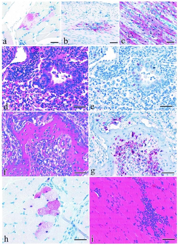
FIG. 2.
Experimental studies of chickens, ducks, and mice inoculated with DK/Anyang/AVL-1/01. Photomicrographs of hematoxylin-and-eosin-stained tissue sections (d, f, and i) or sections stained by IHC methods to demonstrate AIV (a to c, e, g, and h). (a) AIV antigen in cytoplasm of skeletal muscle fibers from a 4-week-old chicken that died 3 days after i.n. inoculation. Bar = 18 μm. (b) AIV antigen in cytoplasm and nuclei of smooth muscle fibers within the tunica muscularis of duodenum from a 4-week-old chicken that died 3 days after i.n. inoculation. Bar = 40 μm. (c) AIV antigen in cytoplasm and nuclei of cardiac muscle fibers from a 4-week-old chicken that died 2 days after i.n. inoculation. Bar = 40 μm. (f) Focal acute periosteal necrosis in pneumatic bone of the cranium in a 2-week-old duck euthanatized 2 days after i.n. inoculation. Bar = 35 μm. (g) AIV antigen in periosteal mesenchymal cells in a 2-week-old duck euthanatized 2 days after i.n. inoculation. Bar = 35 μm. (h) AIV antigen in perilaryngeal skeletal myocytes in a 2-week-old duck euthanatized 2 days after i.n inoculation with DK/Anyang/AVL-1/01 virus. Bar = 15 μm. (i) Focal myofiber degeneration with corresponding lymphohistiocytic myositis in perilaryngeal skeletal muscle in a 2-week-old duck euthanatized 7 days after i.n. inoculation. Bar = 35 μm. (d) Necrotizing bronchitis with neutrophilic inflammation and associated lymphohistiocytic alveolitis in a 4-week-old BALB/c mouse that was euthanatized 4 days after i.n. inoculation. Bar = 35 μm. (e) AIV antigen in nuclei of bronchial epithelium and type II pneumocytes in 4-week-old BALB/c mice that was euthanatized 4 days after i.n. inoculation. Bar = 35 μm.

