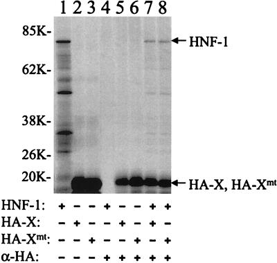
FIG. 1.
Coimmunoprecipitation of HNF-1 with X and Xmt proteins in vitro. HNF-1, HA-X, and HA-Xmt were labeled with [35S]methionine and synthesized in vitro with rabbit reticulocyte lysates. Details of the procedures are described in Materials and Methods. They were then subjected to gel electrophoresis without immunoprecipitation (lanes 1 to 3) or after immunoprecipitation (lanes 4 to 8) with the anti-HA antibody. Lane 1, HNF-1 without immunoprecipitation; lane 2, HA-X without immunoprecipitation; lane 3, HA-Xmt without immunoprecipitation; lane 4, HNF-1 with immunoprecipitation with the anti-HA antibody; lane 5, HA-X with immunoprecipitation; lane 6, HA-Xmt with immunoprecipitation; lane 7, HA-X and HNF-1 immunoprecipitated with the anti-HA antibody; and lane 8, HA-Xmt and HNF-1 immunoprecipitated with the anti-HA antibody. The arrows mark the locations of the HNF-1, HA-X, and HA-Xmt bands. In lane 1, protein bands smaller than the size of HNF-1 were also detected. The nature of these bands is unclear. They might be degraded HNF-1 or N-terminally truncated HNF-1 synthesized from the internal ATG codons. Sizes are shown on the left in kilodaltons.

